A cloak of the brain , a mantle of the brain , or a cloak of the cerebral hemispheres , a mantle of the cerebral hemispheres , pallium , pallium or mantellum in the anatomy of the brain is a set of layers of gray and white matter covering the cerebral hemispheres in lower chordates . In mammals, a homologous structure, which reaches significant development in them, is called the cerebral cortex .
| Cloak of the brain, or pallium, of chordate animals , it is also the cerebral cortex in mammals and humans | |
|---|---|
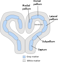 Schematic section of the forebrain of a lower chordal animal , such as a shark . | |
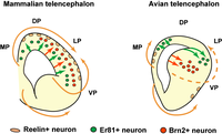 Schematic illustration of the difference in specialization and migration of cloak neurons during embryonic development in mammals and birds . | |
| Part | end brain |
| Components | ancient bark , old bark and new bark |
| Catalogs | |
The term mantellum , or the mantle of the brain , usually applies only to the evolutionarily oldest and most primitive, thin, two-layer version of this structure found in cyclostomes . More developed versions of this structure in bony fishes and other vertebrates , consisting of three layers of cells, are commonly called pallium or brain cloak , and five-layer or six-layer structures characteristic of mammals are called the cerebral cortex.
The part of the terminal brain that is located under the brain cloak, cerebral cortex or mantle of the brain is called the subpallium, submantellum, or, respectively, podplaschevye or submantium structures (in mammals, respectively, subcortical or subcortical structures). In the lower chordates, the cloak or mantle of the brain is a relatively simple structure consisting of only three layers of cells (and in cyclostomes, as already mentioned, even of only two layers), in which only 3-4 different regions or regions are histologically and neurochemically distinguished, plus olfactory bulb . It was previously assumed that the cape or mantle of the brain in the lower chordates is homologous to the cerebral cortex in mammals, and the structures of the terminal brain in the lower chordates that are directly under the cloak or mantle in the lower chordates are homologous to the basal nuclei in mammals. However, from the data of comparative molecular genetic studies, as well as the study of embryogenesis, it follows that from the pallium of the ancient common ancestors of reptiles and mammals during phylogenetic evolution both structures developed in living mammals and belong to the cerebral hemispheres (allo-cortex and isocortex) and part of the structures in living mammals are classified as basal ganglia, namely, the so-called pallial, or mantle, nuclei - the fence and tonsil . In the course of phylogenetic evolution, all the other basal nuclei developed from the subglacial structures, in particular, the striatum , the pale ball , the shell , the lenticular nucleus , the diagonal-nameless nucleus, the preoptic nucleus , as well as the corticoid structures of the olfactory tubercle . [one]
In mammals, both the cortical and now subcortical parts of the brain cloak have undergone significant evolutionary complication. From the cortical part of the cloak of the brain, the cerebral cortex developed, for the most part consisting of progressively more complex, thickening and expanding with the formation of convolutions, in the evolutionary direction from lower mammals to primates and humans, a five- and then six-layer structure called the isocortex , or “typical bark ”, And to a lesser extent, bordering the isocortex, from a thinner and more primitively arranged three- or even two-layer structure called the allocortex , or“ atypical bark ”. Allocortex, in turn, is subdivided into medially (medially) located hippocampal allocortex, and laterally (laterally) located olfactory cortex , including its most rostral (most anterior) part - olfactory bulb and anterior olfactory cortical fields. Thus, allocortex is a significant part of the olfactory brain , and vice versa - the olfactory brain consists mainly of allocortex.
Content
Anatomical structure
Evolution
The phylogenetic evolution of the dorsal (upper) part of the pallium, or brain cloak, has not been fully elucidated. Some authors believe that the brain cloak of the once-existing ancient lower amniotes (common ancestors of reptiles and mammals) in mammals basically turned into hippocampal allocortical and parahippocampal (near-hippocampal) mesocortical transitional areas, preserving the character, having a distinctive character. At the same time, the five- or six-layer structure of the isocortex or neocortex, characteristic only for mammals, according to the opinion of these authors, gradually arose during the evolution of mammals as a de novo growth, on top of the existing pallic (cloak) brain structures, and not as a result of their evolutionary complication . The third authors believe that different parts of the cerebral cortex of mammals were formed from different parts of the brain cloak of the most ancient common ancestors of reptiles and mammals: the medial (median) part of the dorsal pallium of these ancestors gave rise to allotexter, and the lateral (lateral) parts of the dorsal pallium and lateral pallium caused the beginning of the isocortex. [2]
In fish and amphibians (i.e. anamniotic chordates)
Among amphibian structures, the medial, dorsal, lateral and ventral parts of the cape of the brain (pallium), as well as the striatal, pallidary, diagonal-anonymous and preoptic parts of the subpallium, or the system of basal nuclei are well distinguished among the structures of the end brain. However, the pallial part (cloak) of the final brain in them does not show a clearly expressed, visible in an optical microscope with the usual Nissl staining methods, that is, a histologically distinguishable three-layer structure (for its detection, immunohistochemical staining is necessary). The amphibian brain cloak consists of a mixture of glutamatergic (stimulating) and GABAergic (inhibitory) neurons. Subpallium, or basal nuclei, in them mainly consists of GABAergic inhibitory neurons.
The structure of the cloak of the brain in amphibians is generally similar to that of fish. However, even in cartilaginous and especially bony fish, the three-layer structure (in cyclostome fish, two-layer structure) of the structure of the pallium or cloak of the brain is clearly distinguishable histologically using an ordinary light microscope when stained according to Nissl.
In reptiles and birds
In reptiles, the three-layered histological structure of the medial (median) and dorsal (upper) parts of the brain cloak is clearly expressed. Such a morphological structure of the cloak of their brain is very similar to allocortex (or rather paleocortex) in mammals. Due to this similarity, the terms “allocortex” or “paleocortex” are often applied to reptiles that do not yet have the isocortex or neocortex (the five- or six-layer structure of the cerebral cortex that is available only in mammals).
The lateral (lateral) and ventral (lower) parts of the cloak of the brain or pallium of reptiles, however, have a hypopallic bilayer structure, and are called hypopallium . This part of their pallium consists of a superficial olfactory cortex , covering itself with deep pallial nuclei (fencing and amygdala), and resembles in its two-layer structure the mantle of the brain of the lower chordates (cyclostomes). The hypopallium region is also called the dorsal supraventricular cushion ( eng. Dorsal ventricular ridge ), in which the anterior claustral part adjacent to the fence and the posterior amygdaloid part covering the amygdala of the brain are isolated.
In birds, the density and total number of nerve cells in pallium (brain cloak) are significantly increased, while maintaining the traditional three-layer morphological structure for reptiles. Such a significantly increased density of neurons in the pallium leads to the apparent disappearance of a clear separation of the layers in the medial (median) and dorsal (upper) parts of the bird's raincoat when viewed in a conventional light microscope with Nissl-stained preparations. However, with immunohistochemical staining, the three-layer structure of the cloak structure of the brain of birds is clearly visible. In the hypopallial region in birds, compared with reptiles, the size and thickness of the olfactory cortex decreased significantly. At the same time, hypopallial nuclei (or the nuclei of the dorsal supraventricular roller) in birds significantly increase in size and differentiate in cellular composition, compared with reptiles.
In mammals and humans
In mammals, especially in higher primates and humans, the structure homologous to pallium (cloak of the brain) of the lower chordates has reached a large development and covers most of the end brain. This happened mainly due to the massive increase in the surface of the isocortex (“typical cortex”), or, in other words, the neocortex (“new cortex”).
Traditionally, the “cloak” (pallium) of the final brain, or rather, the cerebral cortex, in mammals and humans, it was customary to subdivide into archipallium (“ancient cloak” or ancient bark), paleopallium (“old cloak”, or old bark) and neopallium (“new cloak”, or new bark). However, this unit has been considered obsolete since the end of the 20th century. At the same time, it was proposed to subdivide the cloak or cortex of the mammalian cerebral hemispheres into the medial, dorsal, lateral and ventral parts.
Previously it was believed that only the cortex of the cerebral hemispheres of the mammals' brain originated from the ancient common ancestors of reptiles and mammals from pallium (a cloak of the brain), while the basal nuclei of mammals originated from the subpallium (subplacus structures) of these ancestors. However, a more thorough comparative study of molecular genetic markers of different brain regions in mammals and reptiles, as well as details of the process of embryonic brain development, showed that both the cortical structures of mammals (allocortex and isocortex) and some mammalian basal nuclei, namely the so-called "pallial nuclei" (fence and tonsil). And from the subpallium (subplacus structures) of these ancient animals, a striatum, a pale ball, a shell, a lenticular nucleus, diagonal-nameless and preoptic nuclei, as well as the corticoid structures of the olfactory tubercle of mammals originated .
See also
- Bird's Cloak
Notes
- ↑ Fisher, Robin; Xie, Yuan-Yun. Growth Defects in the Dorsal Pallium after Genetically Targeted Ablation of Principal Preplate Neurons and Neuroblasts: A Morphometric Analysis (Eng.) // ASN Neuro : journal. - 2010 .-- 4 October ( vol. 2 , no. 5 ). - P. AN20100022 . - DOI : 10.1042 / AN20100022 . - PMID 20957077 .
- ↑ Butler, Ann B .; Reiner, Anton; Karten, Harvey J. Evolution of the amniote pallium and the origins of mammalian neocortex (Eng.) // Annals of the New York Academy of Sciences : journal. - 2011 .-- April ( vol. 1225 , no. 1 ). - P. 14-27 . - DOI : 10.1111 / j.1749-6632.2011.06006.x . - PMID 21534989 .