Thalamus pillow ( lat. Pulvinar thalami , or simply lat. Pulvinar ) is a group of nuclei (Taurus neurons ) located in the thalamus (part of the diencephalon of vertebrates ) of animals . This group of nuclei is collectively called the pulvinar, or thalamus pillow ( lat. Pulvinar thalami ), and as a collection of individual nuclei, by the pulvar nuclei, or thalamus pillow nuclei ( lat. Nuclei pulvinares thalami ).
| Thalamus pillow | |
|---|---|
 Hindbrain , diencephalon and midbrain , posterior-lateral view. The thalamus pillow is visible near the top of the image. | |
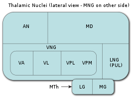 Thalamus nuclei : MNG = Middle group of nuclei AN = Thalamus anterior nuclei MD = Medial Dorsal Core VNG = Ventral nucleus group VA = Ventral anterior core VL = Ventral lateral nucleus VPL = Ventral posterolateral nucleus VPM = Ventral Posteromedial Nucleus LNG = Lateral group of nuclei PUL = Thalamus Pillow MTh = Metatalamus LG = Lateral Cranial MG = Medial Cranial | |
| Part | thalamus |
| Components | anterior , inferior , lateral (lateral) and medial (median) nuclei of the pillow |
| Catalogs | |
| |
The nuclei of the thalamus pillow are usually assigned to the lateral group of nuclei of the thalamus in rodents and in predatory mammals . However, in humans and other higher primates, they are isolated in a separate group of thalamic nuclei.
Content
Anatomical structure
According to the agreement, the group of nuclei of the thalamus pillow is divided into four nuclei:
| Alphanumeric designation for Terminologia Anatomica | Latin name according to Terminologia Anatomica | Name in English | Name in Russian |
|---|---|---|---|
| A14.1.08.611 | nucleus pulvinaris anterior | anterior pulvinar nucleus | Front core pillows |
| A14.1.08.612 | nucleus pulvinaris inferior | inferior pulvinar nucleus | Lower core pillows |
| A14.1.08.613 | nucleus pulvinaris lateralis | lateral pulvinar nucleus | Lateral (lateral) core of the pillow |
| A14.1.08.614 | nucleus pulvinaris medialis | medial pulvinar nucleus | Medial (median) core of the pillow |
These nuclei have the following connections with other parts of the brain :
- The lateral (lateral) and lower nuclei of the thalamic pillow have close connections with the visual cortex of the cerebral hemispheres .
- The dorsal part of the lateral nucleus of the thalamus pillow has close connections with the posterior parietal cortex of the cerebral hemispheres and with the so-called dorsal bundle.
- The medial (median) core of the thalamus pillow has close ties with the cingulate cortex , posterior parietal cortex , premotor and prefrontal cortex of the cerebral hemispheres . [2]
- The thalamus pillow also receives incoming information from the upper hills of the quadruple . These nerve fibers end in the lower , lateral (lateral) and medial (median) nuclei of the thalamus pillow, and, apparently, play a role in maintaining saccadic and anti-saccadic eye movements compensating them, [3] [4] as well as in the regulation of visual attention . [5] [6]
Clinical Importance
Damage to the nuclei of the thalamus pillow can lead to syndromes of neglect of visual signals on the affected side, as well as to impaired concentration of visual attention . [7]
Other animals
The value of the thalamus pillow is different in different species of vertebrates . The thalamus pillow is practically absent as a pronounced thalamic structure in rats and other rodents . And in cats and other carnivorous mammals, the thalamus pillow, due to its small size and weak separation from the lateral group of nuclei , is traditionally combined into a group together with the lateral posterior nucleus , the so-called lateral posterior-pillow group. Meanwhile, in humans, the thalamus pillow is about 40% of the total thalamus volume, which makes it the largest group of thalamic nuclei in humans. [8] Studies on marmoset monkeys show that afferent impulses emanating from the retinal (retinorecipient) region of the lower cushion nucleus (medial region of this nucleus) to the visual cortex of MT are key to the maturation of the visual cortex of MT and the dorsal beam, and can also partially replace neurological deficits arising from damage to the primary visual cortex (region V1). [9] [10] [11]
Etymology
The word "pulvinar" ( English pulvinar ) comes from lat. pulvinus , which literally means "pillow." Just as the cervix (cervix, English cervix ) in English scientific texts is often called simply cervix ( cervix ), despite the fact that its correct Latin name is lat. cervix uteri (cervix), and this does not give rise to any ambiguity, since no other anatomical structure is now called cervix, and the thalamus pillow ( Latin pulvinar thalami , English pulvinar of the thalamus ) is often called simply “pulvinar” ( pulvinar) ), and this also does not give rise to any ambiguity. No other anatomical structure in today's anatomical terminology is called pulvinar. [12] Each of the nuclei of the thalamic pillow has its own set of cortical projections (connections with the cerebral cortex ).
Sources
- ↑ 1 2 Foundational Model of Anatomy
- ↑ Cappe C .; Morel A .; Barone P .; Rouiller EM The thalamocortical projection systems in primate: an anatomical support for multisensory and sensorimotor interplay (English) // Cerebral Cortex: journal. - 2009. - Vol. 19 , no. 9 . - P. 2025-2037 . - DOI : 10.1093 / cercor / bhn228 . - PMID 19150924 .
- ↑ Berman R .; Wurtz R. Signals conveyed in the pulvinar pathway from superior colliculus to cortical area mt. (Eng.) // The Journal of Neuroscience : journal. - 2011. - Vol. 31 , no. 2 . - P. 373-384 . - DOI : 10.1523 / jneurosci . 4738-10-10.2011 . - PMID 21228149 .
- ↑ Robinson D .; Petersen S. Responses of pulvinar neurons to real and self-induced stimulus movement (Eng.) // Brain Research : journal. - 1985. - Vol. 338 , no. 2 . - P. 392-394 . - DOI : 10.1016 / 0006-8993 (85) 90176-3 .
- ↑ Petersen S .; Robinson D .; Morris J. Contributions of the pulvinar to visual spatial attention (English) // Neuropsychologia : journal. - 1987. - Vol. 25 , no. 1 . - P. 97-105 . - DOI : 10.1016 / 0028-3932 (87) 90046-7 .
- ↑ Chalupa, L. (1991). Visual function of the pulvinar. The Neural Basis of Visual Function. CRC Press, Boca Raton, Florida, pp. 140-159.
- ↑ Arend I .; Rafal R .; Ward R. Spatial and temporal deficits are regionally dissociable in patients with pulvinar lesions (English) // Brain : journal. - Oxford University Press , 2008. - Vol. 131 , no. 8 . - P. 2140-2152 . - DOI : 10.1093 / brain / awn135 . - PMID 18669494 .
- ↑ LaBerge, D. (1999). Attention pp. 44-98. In Cognitive science (Handbook of Perception and Cognition, Second Edition), Bly BM, Rumelhart DE. (edits). Academic Press ISBN 978-0-12-601730-4 p. 73
- ↑ Warner CE, Kwan WC, Bourne JA The early maturation of visual cortical area MT is dependent on input from the retinorecipient medial portion of the inferior pulvinar (Eng.) // Journal of Neuroscience : journal. - 2012. - Vol. 32 , no. 48 . - P. 17073-17085 . - DOI : 10.1523 / JNEUROSCI.3269-12.2012 . - PMID 23197701 .
- ↑ Warner CE, Goldshmit Y., Bourne JA Retinal afferents synapse with relay cells targeting the middle temporal area in the pulvinar and lateral geniculate nuclei (Eng.) // Front Neuroanat: journal. - 2010 .-- Vol. 4 . - P. 8 . - DOI : 10.3389 / neuro.05.008.2010 . - PMID 20179789 .
- ↑ Warner CE, Kwan WC, Wright D., Johnston LA, Egan GF, Bourne JA Preservation of vision by the pulvinar following early-life primary visual cortex lesions (English) // Curr Biol : journal. - 2015. - Vol. 25 , no. 4 . - P. 424-434 . - DOI : 10.1016 / j.cub.2014.12.12.028 . - PMID 25601551 .
- ↑ Baud, RH, Latin index of TA98, Terminologia Anatomica version 1998 , < https://www.unifr.ch/ifaa/Public/EntryPage/TA98%20Tree/Alpha/All%20KWIC%20W%20LA.htm >
Additional Images
Thalamus
Deep section of the brain stem . Lateral view.
Deep section of the brain stem . Dorsal view.
Diagram showing the central connections of the optic nerves and optic tracts.
The human brain in the mid-sagittal section, left view.