The human heart is a cone-shaped hollow muscular organ, which receives blood from the venous trunks flowing into it and pumps it into the arteries that are adjacent to the heart. The cavity of the heart is divided into two atria and two ventricles. The left atrium and the left ventricle together form the "arterial heart", so named after the blood passing through it, the right ventricle and the right atrium are combined into a "venous heart", named according to the same principle. The contraction of the heart is called systole, and relaxation is called diastole [B: 1] .
| A heart | |
|---|---|
 Human heart | |
 Cardiac cycle - the work of the heart | |
| System | Circulation |
| Blood supply | Right coronary artery , left coronary artery |
| Venous outflow | large heart vein, medium heart vein, small heart vein, anterior vein of the heart, small vein, posterior vein of the left ventricle, oblique vein of the left atrium |
| Innervation |
|
| Lymph | lower tracheobronchial lymph nodes, anterior mediastinal lymph nodes. |
| Catalogs | |
Heart shape is not the same for different people. It is determined by age, sex, physique, health and other factors. In simplified models, it is described by a sphere, ellipsoids, and intersection figures of an elliptic paraboloid and a triaxial ellipsoid. The measure of elongation (factor) form is the ratio of the largest longitudinal and transverse linear dimensions of the heart. With hypersthenic body type, the ratio is close to unity and asthenic - about 1.5. The length of an adult’s heart varies from 10 to 15 cm (usually 12–13 cm), the width at the base is 8–11 cm (more often 9–10 cm) and the anteroposterior size is 5–8.5 cm (usually 6.5–7 cm) . The average heart mass is 332 g for men (from 274 to 385 g) and 253 g for women (from 203 to 302 g) [B: 2]
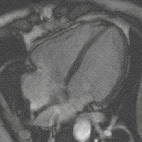
Anatomical structure of the heart
The heart is located in the center of the chest and is shifted by the lower left edge to the left side, in the so-called pericardial bag - the pericardium , which separates the heart from other organs.
In relation to the midline of the body, the heart is located asymmetrically - about 2/3 to the left of it and about 1/3 to the right. Depending on the direction of the projection of the longitudinal axis (from the middle of its base to the apex) onto the anterior chest wall, there is a transverse, oblique and vertical position of the heart. The vertical position is more common in people with a narrow and long rib cage, transverse - in individuals with a wide and short rib cage.
The heart consists of four separate cavities, called chambers: left atrium , right atrium , left ventricle , right ventricle . They are separated by partitions. The right atrium includes hollow, the left atrium - pulmonary veins. The pulmonary artery (pulmonary trunk) and the ascending aorta , respectively, exit from the right ventricle and left ventricle. The right ventricle and the left atrium close the small circle of blood circulation , the left ventricle and the right atrium - a large circle . The heart is located in the lower part of the anterior mediastinum, most of its front surface is covered by the lungs with flowing areas of the hollow and pulmonary veins, as well as the outgoing aorta and the pulmonary trunk. The pericardial cavity contains a small amount of serous fluid. [B: 2]
The wall of the left ventricle is approximately three times thicker than the wall of the right ventricle, since the left must be strong enough to push the blood into the systemic circulation for the whole body (resistance to blood flow in the systemic circulation is several times greater, and the blood pressure is several times higher than in the pulmonary circulation).
There is a need to maintain blood flow in one direction, otherwise the heart could be filled with the same blood that was sent to the arteries before. Responsible for the flow of blood in one direction are the valves, which at the appropriate moment are opened and closed, letting the blood through or putting a barrier to it. The valve between the left atrium and the left ventricle is called the mitral valve or the double valve, since it consists of two lobes. The valve between the right atrium and the right ventricle is called the tricuspid valve - it consists of three lobes. In the heart are still aortic and pulmonary valves . They control the flow of blood from both ventricles.
Blood supply
Every cell of the heart tissue should have a constant supply of oxygen and nutrients. This process is provided by the heart’s own blood circulation through the system of its coronary vessels; it is commonly referred to as “ coronary circulation ”. The name comes from 2 arteries, which, like a crown, braid the heart. The coronary arteries directly extend from the aorta. Up to 20% of the blood pushed out by the heart passes through the coronary system. Only such a powerful portion of oxygen-enriched blood ensures the continuous operation of the life-giving pump of the human body.
Innervation
The heart receives a sensitive, sympathetic and parasympathetic innervation. Sympathetic fibers from the right and left sympathetic trunks, passing in the composition of the heart nerves, transmit impulses that accelerate the heart rhythm, expand the lumen of the coronary arteries, and parasympathetic fibers conduct impulses that slow the heart rhythm and narrow the lumen of the coronary arteries. Sensory fibers from the receptors of the walls of the heart and its vessels go in the composition of the nerves to the corresponding centers of the spinal cord and brain.
Histological structure of the heart
The wall of the heart consists of three layers - the epicardium, myocardium and endocardium. The epicardium consists of a thin (no more than 0.3–0.7 mm) plate of connective tissue, the endocardium consists of epithelial tissue, and the myocardium consists of cardiac striated muscle tissue. Myocardial cells are called cardiomyocytes .
Myocardium is densely riddled with blood vessels and nerve fibers forming several nerve plexuses. For each myocardial capillary, there are about four nerve fibers. [B: 3]
Biophysical view on the structure of the heart
From the point of view of cardiophysics, the heart is a multicomponent polymeric heterogeneous active medium of natural origin. The fine organization of the structure of this environment provides its basic biological functions.
The heterogeneous structure of the heart, which is the basis of its fine organization, was repeatedly confirmed, first using electrophysiology methods and then using computational biology methods (see figure).
The autowave properties of cardiac tissue have been actively studied by both Russian and world science for more than half a century.
A new scientific view of this biological object allows for a new approach to solving the problem of creating an artificial heart: the task is to establish production of an artificial polymer active medium based on modern nanotechnology with a similar autowave function [B: 4] [B: 5] .
Cardiac physiology
Heart activity
Historically taken [B: 1] to allocate the following physiological properties of cardiac tissue:
Automation of the heart is the ability of the heart to rhythmically contract under the influence of impulses originating in it.
The excitability of the heart is the ability of the heart muscle to be excited by various stimuli of a physical or chemical nature, accompanied by changes in the physicochemical properties of the tissue.
Conductivity of the heart - is carried out in the heart electrically due to the formation of the action potential in the cells of pace makers. The place of transition of excitation from one cell to another, are the nexus.
Contractility of the heart - the force of contraction of the heart muscle is directly proportional to the initial length of the muscle fibers.
Myocardial refractoriness is a temporary state of non-irritability of tissues.
The phenomena of automatism, excitability, and conduction can be united by the notion “ autowave function of the heart ” [B: 4] [B: 5] .
It is believed that cardiac activity is aimed at ensuring the pumping function of the heart , that is, "the main physiological function of the heart is the rhythmic injection of blood into the vascular system" [B: 6] .
Blood circulation
Performing a pumping function in the circulatory system, the heart constantly pumps blood into the arteries. The human heart is a kind of pump that provides a continuous and continuous movement of blood through the vessels in the right direction.
Bicuspid and tricuspid valves provide blood flow in one direction - from the atria to the ventricles.
Heart work cycle
A healthy heart rhythmically and without breaks is compressed and unclenched. In one cycle of the heart, there are three phases:
- Blood-filled atria contract. At the same time, blood is pumped through the open valves into the ventricles of the heart (at this time they remain in a state of relaxation). Atrial contraction begins at the site of the inflow of veins into it, so their mouths are compressed and the blood cannot get back into the veins.
- There is a contraction of the ventricles with simultaneous relaxation of the atria. The tricuspid and bicuspid valves that separate the atria from the ventricles rise, slam, and prevent blood from returning to the atria, and the aortic and pulmonary valves open. The contraction of the ventricles injects blood into the aorta and pulmonary artery.
- Pause (diastole) a short period of rest of the body. During a pause, blood from the veins enters the atria and partially flows into the ventricles. When a new cycle begins, the blood remaining in the atria will be pushed into the ventricles - the cycle will be repeated.
One cycle of the heart lasts about 0.85 seconds, of which only 0.11 seconds are needed for the atrial contraction, 0.32 seconds for the ventricular contraction, and the longest rest period, which lasts 0.4 sec. The heart of an adult, who is at rest, works in the system at about 70 cycles per minute.
Heart Automatism
A certain part of the heart muscle specializes in issuing control signals to the rest of the heart in the form of corresponding pulses of autowave nature ; This specialized part of the heart is called the Conductive System of the Heart (PSS). That it provides the automaticity of the heart.
| Automatism - the ability of the heart to be excited under the influence of impulses that occur in cardiomyocytes without external stimuli. Under physiological conditions, ACS has the highest automatism in the heart, therefore it is called an automatic center of the first order.A.V. Ardashev et al., 2009 [B: 7] |
The sinoatrial node , called the 1st order pacemaker located on the fornix of the right atrium, is an important part of the PSS. [1] By sending regular autowave pulses, it controls the frequency of the cardiac cycle . These impulses through the paths of the atria enter the atrioventricular node and then into the individual cells of the working myocardium, causing them to contract.
Thus, the PSA, by coordinating atrial and ventricular contractions, ensures rhythmic cardiac performance, that is, normal cardiac activity .
Heart work regulation
The work of the heart is regulated by the myogenic, nervous and humoral mechanisms.
Myogenic, or hemodynamic, regulation mechanism is divided into: heterometric and homeometric [B: 8] .
The nervous system regulates the frequency and strength of heart contractions: (the sympathetic nervous system causes an increase in contractions, the parasympathetic weakens).
The effect of the endocrine system on the heart occurs through the use of hormones that can strengthen or weaken the strength of heart contractions, and change their frequency. The adrenal glands can be considered the main endocrine gland that regulates the work of the heart: they release the hormones epinephrine and norepinephrine , in addition to them also accelerate heart contractions: serotonin , thyroxin , Ca 2+ whose action on the heart corresponds to the functions of the sympathetic nervous system. Calcium and potassium ions, as well as endorphins and many other biologically active substances also have an effect on the heart. However, there are substances that contribute to the slowing of the heart: acetylcholine , bradykinin , K + .
Instrumental methods for diagnosing heart work
Heart ultrasound
Enough informative method of visualization of the structure, physiological processes, pathologies, and hemodynamics (Doppler). In contrast to the methods based on X-ray technology, it has no radiation exposure. The advantages of the method can also be attributed to the speed of research, security, accessibility.
Electrical phenomena
The work of the heart (like any muscle) is accompanied by electrical phenomena that cause the appearance of an electromagnetic field around a working organ. The electrical activity of the heart can be registered using various methods of electrocardiography , which gives a picture of changes in time of the potential difference on the surface of the human body, or electrophysiological studies of the myocardium, allowing to trace the propagation of excitation waves directly on the endocardium. These methods play an important role in the diagnosis of heart attack and other diseases of the cardiovascular system.
Acoustic phenomena
Acoustic phenomena, called heart tones, can be heard by applying an ear or stethoscope to the chest. Each heart cycle is normally divided into 4 tones. The ear is heard at each contraction of the first 2. Longer and lower is associated with the closing of two-and tricuspid valves, shorter and higher - this closes the valves of the aorta and pulmonary artery. Between one and the second tone is the phase of ventricular contraction .
Mechanical Activity
Heart contractions are accompanied by a number of mechanical manifestations, registering which, you can also get an idea of the dynamics of the contraction of the heart. For example, in the fifth intercostal space on the left, 1 cm inside of the midclavicular line, at the time of contraction of the heart, the apical impulse is felt. During the diastole period, the heart resembles an ellipsoid, the axis of which is directed from top to bottom and from right to left. With the reduction of the ventricles, the shape of the heart approaches the ball, while the longitudinal diameter of the heart decreases and the transverse increases. The compacted myocardium of the left ventricle touches the inner surface of the chest wall. At the same time, the apex of the heart, lowered to the diaphragm during diastole, rises and hits the front wall of the chest cell at the time of systole. All this causes the appearance of the apical impulse. [B: 6]
To analyze the mechanical activity of the heart using a number of special methods.
Kinethocardiography [approx. 1] - method for recording low-frequency vibrations of the chest caused by mechanical activity of the heart; allows you to study the phase structure of the cycle of the left and right ventricles of the heart simultaneously.
Electromyography is a method of electrical recording of the movement of the heart shadow contour on the screen of an x-ray machine [2] . A photocell connected to an oscilloscope is applied to the screen at the edges of the contour of the heart. When the heart moves, the photocell illumination changes, which is recorded by the oscilloscope as a curve. Curves of contraction and relaxation of the heart are obtained.
Ballistocardiography is a method based on the fact that the expulsion of blood from the ventricles and its movement in large vessels cause fluctuations of the whole body, depending on the effects of reactive recoil, similar to those observed during a cannon shot (the name of the method “ballistocardiography” comes from "- throwing projectile). Body displacement curves recorded by a ballistocardiograph and dependent on the work of the heart normally have a characteristic appearance. To register them there are several different methods and devices.
Dynamo - cardiography is a method based on the fact that the movements of the heart in the chest and the movement of blood from the heart to the vessels are accompanied by a shift in the center of gravity of the chest in relation to the surface on which the person lies. [3] The patient is lying on a special table on which a special device with sensors - converters of mechanical quantities into electrical oscillations is mounted. The device is located under the chest of the test. Shifts of the center of gravity are recorded by the oscilloscope in the form of curves. On the dynamo-cardiogram, all phases of the cardiac cycle are noted: atrial systole, periods of stress of the ventricles and the expulsion of blood from them, the protodiastolic period, periods of relaxation and filling of the ventricles with blood.
Phonocardiography is a method for recording heart sounds on a phonocardiogram. If in the left half of the chest, at the level of the IV — V rib of the subject, attach a sensitive microphone connected to an amplifier and an oscilloscope, it is possible to register heart sounds in the form of curves on photo paper. This method is used to diagnose valvular lesions. [3]
See also
- Auscultation
- Cardiology
- Heart biophysics
- Circulation
- Peripheral heart
- Pulse
- Congenital heart defects
- Heart disease
- A heart
- Artificial heart
- The cardiovascular system
- Heart symbol
- Abiocor
Comments
- ↑ See also the study of the mechanical activity of the heart
Notes
- ↑ Babsky E. B. Human Physiology. - 2nd ed. - M: Medicine, 1972. - p. 69.
- ↑ Kositsky GI Human physiology. - 3rd ed. - M: Medicine, 1985. - p. 255.
- ↑ 1 2 Kositsky GI Human physiology. - 3rd ed. - M: Medicine, 1985. - p. 256.
- ↑ 1 2 Dudel Y., Ryuegg Y., Schmidt R. et al. Human Physiology: in 3 volumes. Per. from English = Human Physiology / ed. R. Schmidt and G. Tevs . - 3. - M .: Mir, 2010. - T. 1. - 323 with ill. with. - 1000 copies - ISBN 978-5-03-003834-6 .
- ↑ 1 2 M. M. , Lysenkov N. K. , Bushkovich V. I. Human Anatomy. - 11th revised and enlarged. - M .: Medicine, 1985.
- ↑ Histology. - M.
- ↑ 1 2 Elkin Yu. E. , Moskalenko A.V. Basic mechanisms of cardiac arrhythmias // Clinical arrhythmology / Ed. prof. A. V. Ardashev. - M .: MEDPRAKTIKA-M, 2009. - P. 45-74. - 1220 s. - ISBN 978-5-98803-198-7 .
- ↑ 1 2 Tachycardia as “Shadow Play” // Tachycardia / Takumi Yamada, editor. - Croatia: InTech, 2012. - p. 97-122. - 202 p. - ISBN 978-953-51-0413-1 .
- ↑ 1 2 Human physiology / ed. V.M. Pokrovsky and G.F. Korotko . - 3. - M .: Medicine, 2007. - 656 p. - (Educational literature for medical students). - 10 000 copies - ISBN 5-225-04729-7 .
- ↑ Anatomy and physiology of the cardiac conduction system // Clinical arrhythmology / Ed. prof. A. V. Ardashev. - M .: MEDPRAKTIKA-M, 2009. - P. 35-41. - 1220 s. - ISBN 978-5-98803-198-7 .
- ↑ Sudakov KV Normal physiology. - M .: Medical Information Agency, 2006. - p. 329. - 920 p. - ISBN 5-89481-294-1 .