Rhynchonelliformea (lat.) - subtype of the brachopod (brachiopod).
| Rhynchonelliformea | |||||||||||||||||||||||||||||||||||||||||
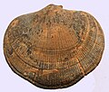
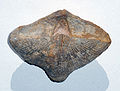
Fossil Shells Rhynchonelliformea | |||||||||||||||||||||||||||||||||||||||||
| Scientific classification | |||||||||||||||||||||||||||||||||||||||||
|---|---|---|---|---|---|---|---|---|---|---|---|---|---|---|---|---|---|---|---|---|---|---|---|---|---|---|---|---|---|---|---|---|---|---|---|---|---|---|---|---|---|
| |||||||||||||||||||||||||||||||||||||||||
| International scientific name | |||||||||||||||||||||||||||||||||||||||||
Rhynchonelliformea A. Williams, Carlson, Brunton, Holmer & Popov, 1996 | |||||||||||||||||||||||||||||||||||||||||
| Classes | |||||||||||||||||||||||||||||||||||||||||
| |||||||||||||||||||||||||||||||||||||||||
| Geochronology appeared 529 million years
◄ Nowadays◄ Cretaceous-Paleogene extinction◄ Triassic extinction◄ Mass Permian Extinction◄ Devonian extinction◄ Ordovician-Silurian extinction◄ Cambrian explosion | |||||||||||||||||||||||||||||||||||||||||


Representatives of this subtype are characterized by a calcite shell consisting of abdominal and dorsal cusps. These flaps are usually articulated through teeth, denticles, or plates. The intestine ends blindly, the anus is absent.
As a rule, these are dioecious animals. After fertilization of the egg, radial fragmentation of the blastomeres begins. Gastrulation is usually done by intussusception, and the mesoderm is formed by the enterocele method. During the development of the larva, the blastopore closes. The larva is not capable of self-feeding. Having lived from several hours to several days, it sinks onto the substrate, attaches to it and undergoes metamorphosis. A secondary mouth forms at the site of the blastopore.
Rhynchonelliformea lead a motionless lifestyle on the seabed. They feed on clusters of bacterial cells and molecules of organic substances, filtering them out of the water.
Representatives of the subtype are widespread in the seas with normal salinity. Different species are found at different depths, but usually no more than 300 m. Rhynchonelliformea have been known since the Atdaban age of the Early Cambrian. They were very numerous and diverse in the geological past.
Study History
Of the genera that make up this group and are currently recognized, the genus Terebratula was the very first to be described. His description was published in 1776 by O. F. Müller [1] .
As an independent subtype, Rhynchonelliformea was first identified in 1996 by Alvin Williams, Sandra J. Carlson, Howard Branton, Lars Holmer, and L. E. Popov [2] . These five authors participated in writing volumes on brachiopods for the new publication of the fundamental work “Treatise on Invertebrate Paleontology” and joined forces to develop a new classification of this group of animals [3] . In 1996, they published an article proposing a new brachiopod system based on data from anatomy, embryology, and molecular biology [2] [3] . In subsequent years, this system was adopted in the Treatise on Invertebrate Paleontology and several other works. However, not all specialists accepted the new classification. G. A. Afanasyeva in 2008 criticized her and suggested using the previous system, arguing at the same time that the question of the systematics of brachiopods remains open [4] .
Structural features
Sink
The shell consists of two cusps - the ventral and dorsal [5] . Its dimensions are usually a few centimeters [6] . The largest shells, reaching a size of about 40 cm, belonged to representatives of the Paleozoic genus Gigantoproductus [6] . Of the modern representatives of the subtype, the largest belong to the species Magellania venosa , the length of their shells reaches 8.4 cm [6] .
The edge of the shell on which the crown is located is called the rear edge, and the opposite edge is called the front edge. In the representatives of the classes Rhynchonellata and Strophomenata, the flaps are usually articulated through the locking joint - two teeth under the crown at the posterior edge of the abdominal cusp, entering the corresponding fossae on the dorsal cusp. In some forms (suborder Productidina ) the castle joint is lost again [7] . The articulation of the valves is absent in many ancient forms - in the order Chileida , as well as some representatives of the class Obolellata [7] . In other Obolellata, articulation was carried out by the teeth of the abdominal cusp and the fossa of the dorsal cusp [7] . Representatives of the order Dictyonellida [8] and the class Kutorginata articulation was carried out by means of grooves on the locking edges of the valves [9] [10] .
The shell is formed of calcite and coated on the outside with a thin organic layer - periostracum . The calcite component is usually two- or three-layer, rarely consists of one layer. The primary (outer) layer is formed by small calcite crystals, often having a needle shape and directed perpendicular to the surface of the shell. The secondary layer has a fibrous (fibrous), or lamellar or cross-lamellar microstructure. The tertiary layer, if developed, consists of polygonal calcite prisms directed perpendicular to the surface.
Musculature
There are muscles of leaflet openers (diductors) and leaflet closers (adductors), as well as leg muscles.
Digestive system
In modern representatives, the intestinal tract ends blindly, the anus is absent [11] . Representatives of the extinct class Kutorginata may have had an anus [12] .
Life cycle
Reproduction
Most modern species are dioecious. However, hermaphroditic species are also found, which differ in general in smaller sizes [13] .
Sexual dimorphism is absent in modern forms, with the exception of Lacazella mediterranea [13] .
Ovum fertilization occurs in the mantle cavity of the female or in the external environment.
In some species (especially in hermaphroditic ones), the development of larvae occurs on the inner surface of the mantle of the parent individual, or within its lophophore. For representatives of the genus Lacazella , brood chambers that form on the lophophore serve for this purpose, and brood chambers in the body cavity for Argyrotheca and Gwynia capsula [14] .
Individual Development
The individual development of Rhynchonelliformea is judged by modern representatives of this group, of which embryology is best studied in Calloria inconspicua , Notosaria nigricans , Terebratulina septentrionalis , Terebratulina retusa and Terebratalia transversa [15] .
Embryo Development
Individual development by the example of Terebratalia transversa
After fertilization of the egg with a sperm, a zygote is formed [15] . Usually it is covered with a membrane of fertilization, which prevents the penetration of other sperm [16] .
The fragmentation of zygotes into blastomere cells is radial [17] . After the fourth phase of fragmentation, in which the number of cells reaches 16, further division often occurs randomly and asynchronously. As a result, a blastula is formed with an internal cavity - blastocele [18] .
Then the blastula turns into gastrula . As with other brachiopods, this transformation ( gastrulation ) usually occurs by intussusception (by digging the blastula wall into a blastocele). The site where the bulging occurs is called the vegetative pole. At the site of invagination, a primary mouth is formed - a blastopore , the invaded part of the wall turns into the endoderm , and the rest - into the ectoderm . However, an exception is also known: in individuals of the Lacazella mediterranea species, gastrulation occurs by delamination (stratification of the blastula into two layers of cells that form the endoderm and ectoderm) [18] .
At the stage of late blastula or early gastrula, cilia develop on the surface of the embryo, and it becomes capable of moving in the water column [18] . At the animal pole of the body [19] (opposite to the vegetative, that is, blastopore), an apical bundle of long fixed cilia is formed, presumably performing sensory functions [18] [20] . At the same time, perhaps, a breakthrough of the fertilization membrane occurs with the embryo coming out, however, this process is poorly studied [18] .
During the further development of gastrula, it acquires an elongated shape. In this case, the rounded blastopore also becomes elongated, and the side of the body on which it is located becomes the abdominal side. The animal pole with the apical bundle of cilia shifts somewhat to one of the ends of the blastopore and becomes the front end of the body [19] .
The mesoderm is formed by the enterocele, that is, from the endoderm [18] . The endoderm section on the dorsal side thickens and gives rise to a layer of mesoderm cells. Growing in the lateral directions, this layer forms coelomic cavities [21] . Messages on the formation of mesoderm in some representatives by the schizocele method (from cells at the border of the ectoderm and endoderm) have not been confirmed [18] .
After the appearance of the mesoderm, the blastopore gradually disappears, overgrowing from the posterior end to the anterior [21] . Its final extinction is regarded as the transformation of an embryo into a larva [15] .
Larval Development
The formed larva is lecithotrophic, that is, it is unable to feed [22] . The body of the larva is transversely divided into anterior, trunk and posterior (foot) sections [21] . In the anterior part there are sometimes paired eyes, which in different species have from a few to a few dozen [22] . Some representatives along the posterior edge of the anterior section have developed several tens of granules, called vesicular bodies. The functions of these bodies are unclear [20] . The trunk section forms a fold that spans the back section. Often at the edge of this fold there are 4 bundles of bristles - two lateral and two dorsal [23] . These bristles perform a tactile function and also serve as a means of protection [24] . In mature larvae, cilia are preserved mainly in the anterior part; moreover, in many species, a band of motor cilia is developed along the perimeter of the anterior part; in addition, cilia remain on the abdominal side of the trunk, where they form a longitudinal strip [20] .
Swimming of the larva lasts from several hours to several days, after which the larva settles to the bottom or other suitable substrate. The posterior region secretes an adhesive or mucous substance and attaches to the substrate [25] .
Further Development
Before the larva settles on the substrate or immediately after that, it loses the apical bundle of cilia and eyes [26] .
In the process of settling or somewhat later, the larva undergoes metamorphosis . The edges of the folds of the trunk rise up and cover the front section, forming a mantle [27] . A shell begins to form on the outer surface of the mantle [26] . Sometimes, shell formation begins even before metamorphosis [28] .
After the larva attaches to the substrate, its posterior part turns into a pedicle [29] .
In place of the former blastopore, a new mouth opening is formed. Lophophore is formed around it from the tissues of the anterior section [26] .
Lifestyle
In adulthood, representatives of Rhynchonelliformea lead an almost immobile lifestyle.
Ecological types
According to the ratio with the substrate, representatives of this group belong to three ecological types [30] .
- An anchor ecological type is the most common. The body is attached to the substrate with the help of a leg that exists throughout the life of the animal. Representatives of this ecological type are characterized by convex flaps. It is subdivided into three ecological subtypes [30] :
- The main subtype of the anchor type. The leg is large, sometimes branched, holds the shell above the substrate without the help of supporting formations.
- Reference subtype. In adult stages, the leg is not able to hold the shell over the substrate and the crown serves as an additional support.
- Complicated subtype. The function of the additional support is performed by a high area - a section of the shell along the posterior edges of the valves.
- Free-lying ecological type. The body freely lies on the substrate, not attaching to it. The hole in the sink to exit the legs overgrows in adults [30] .
- The first subtype includes forms with strongly convex cusps and a massive crown part.
- The second subtype includes forms lying on the bottom on a wide flattened or concave abdominal cusp.
- The third (pontoon) subtype includes forms that are held on the surface of soft soil with the help of shell processes in the form of needles or growth plates.
- Growing ecological type. The body attaches to the substrate by cementing the surface of the abdominal cusp or part of it. There is no foot attachment. Asymmetric abdominal and flattened dorsal valves are characteristic [30] .
Nutrition
Food for the Rhynchonelliformea representatives is mainly aggregates of bacterial cells and molecules of organic substances . Most of them cannot feed on phytoplankton , in particular, diatoms , which other brachopods feed on [31] .
Power is provided by filtration. Sash flaps are ajar during feeding. The lophophore tentacles closed with each other divide the mantle cavity into two parts: water enters one of these parts from the external environment, then passes through the lophophore and the filtered one leaves the second part of the mantle cavity [32] . The incoming and outgoing water currents are created by the oscillatory movements of the cilia on the tentacles of the lophophore [33] .
Distribution and habitat
Representatives of the subtype are widespread in the seas with normal salinity (at least 26-30 ‰). Most modern species are confined to depths of less than 300 meters, but some species are also found at a depth of 6 kilometers.
In the fossil state, they have been known since the Atdabanian era of the Early Cambrian , they are very numerous and diverse in the Paleozoic sediments, however, by the Mesozoic this diversity has decreased markedly and so far only three groups have survived.
Classification
A. Williams and his coauthors, who identified the subtype of Rhynchonelliformea , included in its composition groups that had previously been united under the name of castle brachiopods ( Articulata ). Castle brachiopods were divided into three independent classes: Rhynchonellata , Strophomenata and Chileata .
In addition, Rhynchonelliformea as an independent class of Obolellata [3] included some forms that were previously included in the castleless brachiopods ( Inarticulata ) [34] .
Another class Kutorginata , also included in this subtype [3] , was previously considered as a Kutorginida order among castleless [35] or as a group of brachiopods of an unclear systematic position [36] .
Thus, five classes are distinguished in the subtype, of which four are completely extinct:
- † Chileata Class
- † Class Obolellata
- † Kutorginata Class
- .. class Rhynchonellata
- † Strophomenata Class
Notes
- ↑ Williams, 1965 , p. H2.
- ↑ 1 2 Williams et al., 1996 .
- ↑ 1 2 3 4 Williams et al., 2000 .
- ↑ Afanasjeva, 2008 , p. 795-796.
- ↑ Likharev, 1960 , p. 116.
- ↑ 1 2 3 Likharev, 1960 , p. 121.
- ↑ 1 2 3 Williams, Brunton & McKinnon, 1997 , p. 364.
- ↑ Holmer, 2000 , p. 198.
- ↑ Williams, Brunton & McKinnon, 1997 , p. 364-365.
- ↑ Popov & Williams, 2000 , p. 208-209.
- ↑ Smirnova, 1990 , p. sixteen.
- ↑ Popov & Williams, 2000 , p. 210.
- ↑ 1 2 Williams et al., 1997 , pp. 125-126.
- ↑ Williams et al., 1997 , pp. 176-178.
- ↑ 1 2 3 Williams et al., 1997 , p. 153.
- ↑ Williams et al., 1997 , p. 156.
- ↑ Smirnova, 1990 , p. 9.
- ↑ 1 2 3 4 5 6 7 Williams et al., 1997 , p. 163.
- ↑ 1 2 Freeman, 2003 , p. 272.
- ↑ 1 2 3 Pennington, Stricker, 2001 , p. 443.
- ↑ 1 2 3 Williams et al., 1997 , p. 165.
- ↑ 1 2 Williams et al., 1997 , p. 173.
- ↑ Williams et al., 1997 , p. 174.
- ↑ Williams et al., 1997 , p. 176.
- ↑ Williams et al., 1997 , p. 179-181.
- ↑ 1 2 3 Williams et al., 1997 , p. 186.
- ↑ Williams et al., 1997 , p. 182.
- ↑ Williams et al., 1997 , p. 183.
- ↑ Smirnova, 1990 , p. eleven.
- ↑ 1 2 3 4 Smirnova, 1990 , p. 30–33.
- ↑ Smirnova, 1990 , p. 34.
- ↑ Abrikosov, 1987 , p. 417.
- ↑ Abrikosov, 1987 , p. 416.
- ↑ Rowell, 1965b , p. 291H
- ↑ Smirnova, 1990 , p. 42.
- ↑ Rowell, 1965a , p. 296H
Literature
- Abrikosov G.G. Class Shoulder-footed (Brachiopoda) // Life of animals. - Moscow: Education, 1987. - T. 1. - S. 415-419. - 300,000 copies.
- Likharev B.K. General part // Fundamentals of paleontology / Editor-in-chief Yu. A. Orlov. The responsible editor of the volume is T. G. Sarycheva. - Moscow: Publishing House of the USSR Academy of Sciences, 1960. - T. Mshanki, brachiopods. Application - phoronids. - S. 115-162. - 344 p. - 3200 copies.
- Smirnova T.N. Brachiopods. - Moscow: Publishing house of Moscow University, 1990. - 72 p. - 500 copies. - ISBN 5-211-02389-7 .
- Afanasjeva GA Supraordinal Brachiopod Classification (English) // Paleontological Journal: Journal. - 2008. - Vol. 42, no. 8 . - P. 792-802 . - DOI : 10.1134 / S0031030108080030 .
- Gary Freeman. Regional specification during embryogenesis in Rhynchonelliform brachiopods (Eng.) // Developmental Biology: Journal. - 2003. - Vol. 261, no. 1 . - P. 268-287. - DOI : 10.1016 / S0012-1606 (03) 00307-5 .
- Lars E. Holmer. Dictyonellida // Treatise on Invertebrate Paleontology. Part H, Brachiopoda / Roger L. Kaesler, Editor. - Revised. - Kansas: The Geological Society of America and the University of Kansas, 2000. - Vol. 2: Linguliformea, Craniiformea, and Rhynchonelliformea (part). - P. 196-200. - ISBN 0-8137-3108-9.
- JT Pennington & SA Stricker. Phylum Brachiopoda // Atlas of Marine Invertebrate Larval Forms / CM Young, Editor. - New York: Academic Press, 2001. - P. 441-461. - ISBN 0127731415 .
- Leonid E. Popov & Alwyn Williams. Kutorginata // Treatise on Invertebrate Paleontology. Part H, Brachiopoda / Roger L. Kaesler, Editor. - Revised. - Kansas: The Geological Society of America and the University of Kansas, 2000. - Vol. 2: Linguliformea, Craniiformea, and Rhynchonelliformea (part). - P. 208-215. - ISBN 0-8137-3108-9.
- AJ Rowell. Class Uncertain // Treatise on Invertebrate Paleontology. Part H, Brachiopoda / Raymond C. Moore, Editor. - The Geological Society of America and the University of Kansas, 1965. - Vol. 1. - P. H296 — H297. - ISBN 0-8137-3008-2 .
- AJ Rowell. Inarticulata // Treatise on Invertebrate Paleontology. Part H, Brachiopoda / Raymond C. Moore, Editor. - The Geological Society of America and the University of Kansas, 1965. - Vol. 1. - P. H260 — H296. - ISBN 0-8137-3008-2 .
- Alwyn Williams. Introduction // Treatise on Invertebrate Paleontology. Part H, Brachiopoda / Raymond C. Moore, Editor. - The Geological Society of America and the University of Kansas, 1965. - Vol. 1. - P. H1 — H5. - ISBN 0-8137-3008-2 .
- Alwyn Williams, C. Howard C. Brunton & David I. McKinnon. Morphology // Treatise on Invertebrate Paleontology. Part H, Brachiopoda / Roger L. Kaesler, Editor. - Revised. - Kansas: The Geological Society of America and the University of Kansas, 1997. - Vol. 1: Introduction. - P. 321-422. - xx + 539 p. - ISBN 0-8137-3108-9 .
- Alwyn Williams, SJ Carlson, CHC Brunton, LE Holmer & LE Popov. A supra-ordinal classification of the Brachiopoda // Philosophical Transactions of the Royal Society of London (series B). - 1996. - Vol. 351. - P. 1171-1193. - DOI : 10.1098 / rstb.1996.0101 .
- Alwyn Williams, MA James, CC Emig, Sarah Mackay & MC Rhodes. Anatomy // Treatise on Invertebrate Paleontology. Part H, Brachiopoda / Roger L. Kaesler, Editor. - Revised. - Kansas: The Geological Society of America and the University of Kansas, 1997. - Vol. 1: Introduction. - P. 7-188. - xx + 539 p. - ISBN 0-8137-3108-9 .
- Alwyn Williams, Sandra J. Carlson & C. Howard C. Brunton. Brachiopod Classification // Treatise on Invertebrate Paleontology. Part H, Brachiopoda / Roger L. Kaesler, Editor. - Revised. - Kansas: The Geological Society of America and the University of Kansas, 2000. - Vol. 2: Linguliformea, Craniiformea, and Rhynchonelliformea (part). - P. 1-21. - ISBN 0-8137-3108-9.