Hemangioblastoma ( other Greek αἷμα - “blood” + ἀγγεῖον - “vessel” + βλαστός - “embryo” + -ωμα - contraction from ὄγκωμα “tumor”) - a tumor of the first degree malignancy of unclear histological origin that occurs within the central nervous system . The most characteristic site of localization is the posterior cranial fossa [1] [2] [3] . It can occur sporadically or in combination with neoplasms of the internal organs, which is characteristic of hereditary Hippel – Lindau disease [4] .
| Hemangioblastoma | |
|---|---|
 Cerebellar hemangioblastoma | |
| ICD-10 | Q 85.8 |
| ICD-10-KM | and |
| ICD-O | M 9161/1 |
| Diseasesdb | 31512 |
| eMedicine | med / 2991 |
| Mesh | D018325 |
Clinically, in most cases, it manifests itself as symptoms of impaired cerebrospinal fluid outflow and / or cerebellar damage. The prognosis for sporadic hemangioblastomas is positive, they do not occur again after they are completely removed by surgery. In patients with Hippel-Lindau disease, the prognosis is worse: even complete removal of the lesion does not prevent the emergence of new tumors of this type in other parts of the central nervous system [5] .
Content
- 1 Study History
- 2 Epidemiology
- 3 Pathological anatomy
- 4 Clinical picture
- 4.1 Clinical picture of cerebrospinal fluid outflow disorders
- 4.2 Clinical presentation of cerebellar lesions
- 4.2.1 Damage to the cerebellar worm
- 4.2.2 Damage to the cerebellar hemispheres
- 4.2.2.1 Lack of coordination of movements
- 4.2.2.2 Muscular hypotension
- 4.2.2.3 Asynergy
- 4.3 Clinical picture of spinal hemangioblastoma
- 5 Diagnostics
- 5.1 Hemangioblastomas of the brain
- 5.2 Hemangioblastomas in the spinal canal
- 6 Differential diagnosis
- 7 Treatment
- 7.1 Surgical treatment
- 7.2 Radiation therapy
- 8 Forecast
- 9 notes
Study History
The study of hemangioblastomas is largely associated with studies of a hereditary disease - Hippel-Lindau disease . German ophthalmologist Eugene von Hippel in 1904 he described changes in the retina characteristic of the disease [6] . In 1926, the Swedish pathologist Arvid Lindau described the characteristic changes in the central nervous system - hemangioblastomas of the cerebellum and spinal cord [2] . A. Lindau, at the autopsy of 50 patients with changes in the retina characteristic of the disease described by Hippel, found characteristic changes in the cerebellum in 10 of them. In two patients, they became the direct cause of death [2] .
In 1927, Lindau made a literature review of 15 observations of “angiomatosis of the nervous system”. In this work, he described the characteristic changes in the internal organs of a disease. After the classical works of the Swedish scientist, whose name began to be called pathology, other publications began to appear, which, citing Lindau, described similar clinical observations [2] .

In particular, in 1928, the founder of American and world neurosurgery H. Cushing described 11 patients operated on for cerebellar hemangioblastomas [2] . Of these, five died either immediately after the operation, or a short time later - high mortality after neurosurgical operations was characteristic of the described time [7] ). After reviewing the work of Lindau, Cushing conducted an additional examination of 6 patients. In one of them, changes described by Hippel and Lindau were found on the retina . In addition, Cushing showed the hereditary nature of the process. Also analyzing his observation and the cases described by Lindau, he noted that the first clinical manifestations of the disease occur in adulthood and are absent in children [2] .
In his work, Cushing concluded that when characteristic changes in the retina are detected, attention should be paid to the presence or absence of symptoms of cerebellar damage, since treatment will have better results in the early stages of the development of the disease [2] .
Epidemiology
Hemangioblastomas account for about 2% of all intracranial neoplasms and about 10% of tumors of the posterior cranial fossa [8] [9] . Their share among intramedullary tumors of the spinal cord is from 2 to 3% [10] [11] [12] . Very rarely observed in the area of the cone and roots of the spinal cord, the terminal thread and peripheral nerves [13] [14] [15] [16] . In a quarter of observations, this pathology is one of the manifestations of Hippel's disease - Lindau. It should be emphasized that in some cases, patients with these tumors do not undergo appropriate screening for the determination of Hippel-Lindau disease. Accordingly, the proportion of hemangioblast associated with it may be greater [8] [9] .
Sporadic hemangioblastomas more often occur in patients at 40-50 years of age, while those associated with Hippel-Lindau disease at 20-30 years of age [1] [3] . Men get sick more often than women - 1.3-2: 1 [8] [17] .
Pathological Anatomy
Macroscopically, these neoplasms are represented by two options [18] :
- Soft tissue tumors that look like a dark cherry encapsulated node
- Large smooth-walled cyst with yellowish transparent contents, on one of the walls of which there is a small tumor node
Rarely enough, necrosis and hemorrhages are found in the tumor tissue [18] .
Microscopically, hemangioblastomas are clusters of thin-walled vessels of various sizes. Interstitial cells with lipid- rich light cytoplasm are located in the intervascular spaces [18] .
For 2010, there are no reports of malignancy of this type of tumor. Histologically, the main symptom of hemangioblastoma is the presence of many capillary channels, which are lined with a single-layered epithelium and surrounded by reticular fibers . Neoplasms consist of three types of cells [19] :
- Endothelial ;
- Pericytes
- Stromal cells are polygonal in shape. Contain fatty inclusions.
According to the histological structure, 3 types of hemangioblastomas are distinguished [19] [20] :
- Juvenile - closely spaced thin-walled elongated capillaries;
- Transitional - thin-walled and stretched capillaries are mixed with stromal cells, some of which are completely filled with fat;
- Pure-cell - a tumor consists of layers of xanthochromic (yellowish) cells located on pathological vessels.
Clinical picture
The characteristic age of patients with the occurrence of hemangioblastoma, not associated with Hippel-Lindau disease, is 40-60 years. With a hereditary disease, these neoplasms occur at a younger age - up to 35 years. The multiplicity of the process indicates the hereditary nature of the disease . In most cases, hemangioblastomas are slowly growing neoplasms of the cerebellum or spinal cord [4] .
The clinical picture of cerebrospinal fluid outflow disorders
Neoplasms in the posterior cranial fossa lead to a violation of the outflow of cerebrospinal fluid in connection with the compression of the cerebrospinal tract - the IV ventricle . The symptom of compression of the cerebrospinal fluid paths and violation of the outflow of cerebrospinal fluid from the place of its production ( lateral ventricles ) is the forced position of the head. In some, it is a consequence of the reflex tension of the neck muscles , in others it is a conscious giving of the head a position in which the outflow of cerebrospinal fluid improves and headaches decrease accordingly [21] .
Headache occurs due to increased intracranial pressure - intracranial hypertension . In the initial stages of pain, they have an incidental character with more or less prolonged intervals. A headache attack can occur without any apparent reason, but more often occurs after physical exertion, coughing, defecation, a quick change in body position in space, as well as in some positions of the head (more often when it is thrown back) [21] .
Vomiting is a very common symptom of the disease. Usually it occurs against the background of a headache attack caused by an increase in intracranial pressure. Vomiting may also occur as an isolated symptom that is independent of headache. In such cases, it is a focal symptom , as it is a result of direct irritation of the vomiting center . An attack can occur with a sharp turn of the head. The combination of vomiting and systematic dizziness indicates damage to the vestibular nuclei bottom of the IV ventricle [21] .
Relatively less common is systematic dizziness. Characterized by the occurrence of dizziness and vomiting with a sharp turn of the head or a quick change in body position - Bruns syndrome . In a number of patients, dizziness is the first symptom of the disease [21] .
An important, albeit non-specific, symptom of tumors of the fourth ventricle are attacks of prolonged muscle tension, or decerebral rigidity . Suddenly, sometimes accompanied by a cry, tonic tension of the muscles of the limbs, trunk and neck occurs. The hand is clenched into a fist, the forearms are slightly bent, the head is thrown back, the back is concave. Convulsions do not go into clonic . The attack lasts 2-3 minutes. Involuntary urination and occlusion of the tongue are usually not observed [21] .
Mental disorders (lethargy, irritability, stupor, etc.) occur in the advanced stages of the disease. The tumor does not affect the centers that are responsible for the psyche. However, the prolonged existence of increased intracranial pressure can lead to the appearance of the above symptoms [21] .
The clinical picture of the disease, the nature and duration of the course are mainly determined by the section in which the IV ventricle is located and where its growth is directed. Death usually occurs either due to paralysis of the respiratory center, due to its compression, or due to the development of brain dislocation - displacement of the tonsils of the cerebellum into the occipital foramen [21] .
Clinical picture of cerebellar lesion
Disorders of statics and coordination of movements , as well as muscle hypotension are characteristic of cerebellar damage.
Cerebellar Worm Damage
The defeat of the cerebellar worm leads to a violation of the statics of the body - the ability to maintain a stable position of its center of gravity, providing stability. With a disorder of this function, static ataxia occurs ( dr. Greek. Ἀταξία - disorder). A person becomes unstable, therefore, in a standing position, he seeks to widely spread his legs, to balance the position of the body with his hands. Static ataxia is especially pronounced in the Romberg position . The patient is invited to stand up, firmly moving his feet, slightly raise his head and stretch his arms forward. In the presence of cerebellar disorders, the patient in this position is unstable, his body sways, and he may fall. In case of damage to the worm, the cerebellum of the patient usually sways from side to side and often falls backward, with pathology of the hemisphere, the cerebellum tends to lean toward the pathological focus. If the static disorder is mild, it is easier to identify in the so-called complicated or sensitized Romberg pose . In this case, the patient is invited to put the feet in one line so that the toe of one foot rests on the heel of the other. The stability estimate is the same as in the usual Romberg pose [22] [23] .
Normally, when a person is standing, the muscles of his legs are tense ( support reaction ), with the threat of falling to the side, his leg on this side moves in the same direction, and the other leg comes off the floor ( jump reaction ). When the cerebellum, mainly its worm, is damaged, the reaction of the support and the jump is disturbed in the patient. Violation of the reaction of the support is manifested by instability in a standing position, especially if his legs are thus closely shifted. Violation of the jump reaction leads to the fact that if the doctor, standing behind the patient and insuring him, pushes the patient in one direction or another, the latter falls with a small push ( symptom of pushing ) [22] .
A gait in a patient with cerebellar pathology is very characteristic and is called "cerebellar". Due to the instability of the body, the patient is uncertain, spreading his legs wide, he is “thrown” from side to side, and when the hemisphere is damaged, the cerebellum deviates when walking from a given direction towards the pathological focus. Especially distinct instability when cornering. While walking, the body of the person is excessively straightened ( Tom's symptom ). The gait in people with cerebellar lesions is much like the gait of a drunk person [22] .
If static ataxia is pronounced, then patients completely lose their ability to control their body and cannot not only walk and stand, but even sit [22] .
Cerebellar Hemisphere
A predominant lesion of the cerebellar hemispheres leads to a disorder of its counter-inertia effects and, in particular, to the appearance of a dynamic disturbance in the coordination of movements , decreased muscle tone and the appearance of asynergies [24] .
Misalignment
It is manifested by the awkwardness of the movements of the limbs, which is especially pronounced during movements requiring accuracy. To identify dynamic ataxia, a number of coordination tests are performed [22] .
- Test for diadochokinesis - the patient is invited to close his eyes, stretch his arms forward and quickly, rhythmically supine and penetrate (rotate outward and inward) the hands. In case of damage to the hemisphere of the cerebellum, the movements of the brush on the side of the pathological process turn out to be more sweeping, as a result, this brush begins to lag. Then they talk about the presence of adiadochokinesis [22] [23] [24] : 359 .
- Finger-nasal test - a patient with closed eyes takes his hand away, and then with his index finger tries to get into the tip of his nose. In the case of cerebellar pathology, the arm on the side of the pathological focus makes an excess movement in volume, as a result of which the patient misses. Intentional tremor (trembling of the fingers) characteristic of cerebellar pathology is also revealed, the severity of which increases as the finger approaches the target [22] [23] [24] : 358 .
- Heel-knee test - a patient lying on his back with his eyes closed raises his leg high and tries to hit the heel of the other leg with his heel. With cerebellar pathology, oversight is noted, especially when performing a homolateral (on the same side) test of the affected cerebellar hemisphere with the foot. If, nevertheless, the heel reaches the knee, then it is proposed to hold it, slightly touching the lower leg, along the tibial crest down to the ankle joint. Moreover, in the case of cerebellar pathology, the heel all the time slides in one direction or the other [22] [23] [24] : 358 .
- Index (finger-finger) test - the patient is invited to get with the index finger to the tip of the explorer’s finger directed at him. In the case of cerebellar pathology, mimicking is noted. The patient’s finger usually deviates toward the affected cerebellar hemisphere [22] [24] : 358 .
- Symptom of Tom-Zhumeni - capturing the subject, the patient disproportionately spreads his fingers wide [22] [24] : 359 .
- “Sample with a cup” - a patient holding a glass of water in his hand splashes water.
- Nystagmus - twitching of the eyeballs when looking to the sides or up. With damage to the cerebellum, nystagmus is considered as the result of intentional trembling of the eyeballs. In this case, the plane of nystagmus coincides with the plane of voluntary eye movements — when viewed from the side, the nystagmus is horizontal, and when viewed upward, it is vertical [22] .The lower line is an attempt by the patient to reproduce the upper
- Speech disorder - occurs as a result of impaired coordination of the muscles that make up the speech-motor apparatus. Speech is slowed down (bradylalia), its smoothness is lost. It acquires an explosive, chanted character (the stress is not placed within the meaning, but at regular intervals) [22] [23] [24] : 359 .
- Handwriting changes - the patient’s handwriting becomes uneven, the letters are distorted, excessively large ( megalography ) [22] [23] .
- Симптом Стюарта-Холмса (симптом отсутствия обратного толчка [23] ) — исследующий просит больного сгибать супинированное предплечье и в то же время, взяв его руку за запястье, оказывает сопротивление этому движению. Если исследующий при этом неожиданно отпустит руку больного, то больной не сможет вовремя притормозить дальнейшее сгибание руки, и она, сгибаясь по инерции, с силой ударит его в грудь [22] [24] :359 .
- Пронаторный феномен — больному предлагается удерживать вытянутые вперёд руки ладонями вверх. При этом на стороне поражённого полушария мозжечка происходит спонтанная пронация (поворот ладони внутрь и книзу) [22] [24] :359 .
- Симптом Гоффа-Шильдера — если больной держит руки вытянутыми вперёд, то на стороне патологического очага рука отводится кнаружи.
- Феномен Дойникова (изменение постуральных рефлексов [23] ) — сидящему больному предлагается кисти с разведёнными пальцами положить на свои бёдра вверх ладонями и закрыть глаза. В случае мозжечковой патологии на стороне патологического очага отмечаются спонтанное сгибание пальцев и пронация кисти [22] .
- Проба Шильдера — больному предлагают вытянуть руки вперёд, закрыть глаза, поднять одну руку кверху и опустить её до уровня другой руки, а затем сделать наоборот. При поражении мозжечка больной опустит руку ниже вытянутой [25] .
Мышечная гипотония
Мышечная гипотония выявляется при пассивных движениях, производимых исследующим в различных суставах конечностей больного. Поражение червя мозжечка ведёт обычно к диффузной гипотонии мышц, тогда как при поражении полушария мозжечка снижение мышечного тонуса отмечается на стороне патологического очага [22] .
Маятникообразные рефлексы обусловлены также гипотонией. При исследовании коленного рефлекса в положении сидя со свободно свисающими с кушетки ногами после удара молоточком наблюдается несколько «качательных» движений голени [25] .
Асинергии
Асинергии — выпадение физиологических синергичных (содружественных) движений при сложных двигательных актах [23] [24] :359 .
Наиболее распространены следующие пробы на асинергию:
- Больному, стоящему со сдвинутыми ногами, предлагают перегнуться назад. В норме одновременно с запрокидыванием головы ноги синергично сгибаются в коленных суставах, что позволяет сохранить устойчивость тела. При мозжечковой патологии содружественное движение в коленных суставах отсутствует, и, запрокидывая голову назад, больной сразу же теряет равновесие и падает в том же направлении.
- Больному, стоящему со сдвинутыми ногами, предлагается опереться на ладони врача, который затем неожиданно их убирает. При наличии у больного мозжечковой асинергии он падает вперёд ( симптом Ожеховского ). В норме же происходит лёгкое отклонение корпуса назад или же человек сохраняет неподвижность.
- Больному, лежащему на спине на твёрдой постели без подушки с ногами, раздвинутыми на ширину надплечий, предлагают скрестить руки на груди и затем сесть. Ввиду отсутствия содружественных сокращений ягодичных мышц больной с мозжечковой патологией не может фиксировать ноги и таз к площади опоры, в результате сесть ему не удаётся, при этом ноги больного, отрываясь от постели, поднимаются вверх (асинергия по Бабинскому ) [22] .
Клиническая картина спинальных гемангиобластом
Для гемангиобластом в области позвоночного канала характерна клиническая симптоматика, вызванная поражением спинальных нервов , белого (проводящих путей) и серого вещества спинного мозга. В зависимости от того, какие структуры поражены, у человека могут возникать слабость в конечностях, спастичность , повышение глубоких рефлексов, боль и нарушение функции тазовых органов в виде недержания либо задержки мочи, очень сильные позывы к мочеиспусканию [4] .
Diagnostics
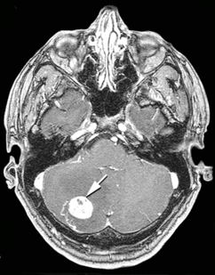
Гемангиобластомы головного мозга
Узел гемангиобластомы состоит из клубка тонкостенных сосудов. При введении в кровь контрастных веществ опухоль накапливает вводимые препараты, благодаря чему хорошо контрастируется на ангиограммах , компьютерных и магнитно-резонансных томограммах. Определяются крупные патологические артерии и вены, могут выявляться артерио-венозные шунты [18] . При проведении компьютерной (КТ) и магнитно-резонансной (МРТ) томографий диагностируются две формы опухоли — узел с или без кистозного компонента. Первая форма характеризуется тем, что большие кисты выполняют большую часть объёмного образования. Узел может вообще не определяться с помощью нейровизуализационных методов исследования [18] .
Киста гемангиобластом обычно округлой или овальной формы, на компьютерной томографии имеет низкую плотность (8—14 единиц Хаунсфилда ). При введении контрастных веществ плотность её содержимого и стенок не изменяется. Узел опухоли определяется на КТ в виде очага повышенной плотности, чаще негомогенной зернистой структуры. Располагается на одной из стенок кисты, вдаваясь в её просвет, хорошо накапливает контрастное вещество [18] .
При кистозной форме опухоли на магнитно-резонансных томограммах хорошо определяется кистозный компонент, который характеризуется низкой интенсивностью сигнала на Т1- и высоким сигналом на Т2-взвешенных томограммах. На этом фоне хорошо визуализируется пристеночно расположенный солидный узел гемангиобластомы, хорошо накапливающий контрастное вещество [18] .
При солидной форме гемангиобластомы в строме новообразования отмечается наличие округлых и извитых участков потери сигнала за счёт кровотока в крупных сосудах опухоли [18] .
Гемангиобластомы в области позвоночного канала
Гемангиобластомы в области позвоночного канала обычно располагаются интрамедуллярно (в спинном мозге), но могут находиться и экстрамедуллярно (вне спинного мозга). В 50 % они поражают грудной отдел и в 40 % шейный. Так как гемангиобластомы представляют собой богатоваскуляризированные новообразования, то их клиническая манифестация может дебютировать субарахноидальным кровоизлиянием [12] [26] .
В большинстве случаев гемангиобластомы в области позвоночного канала представляют собой солитарное образование, однако в 20 % могут наблюдаться множественные новообразования (как правило, при болезни Гиппеля — Линдау). Приблизительно в половине случаев (43—60 %) образуются сирингомиелические полости. Кисты могут достигать больших размеров, располагаясь выше и ниже солидного узла опухоли [26] [27] [28] .
При спинальной ангиографии определяется богато васкуляризированный узел новообразования с крупными приводящими артериями. В связи с этим целесообразным является при подозрении на гемангиобластому проведение селективной ангиографии. Катетеризация артерий, которые кровоснабжают опухоль, может быть использована для одновременной предоперационной эмболизации приводящих сосудов [26] .
Компьютерная томография с контрастным усилением позволяет хорошо визуализировать гемангиобластому. На МР томограммах узел опухоли имеет гипо- или изоинтенсивный сигнал на Т1-взвешенных томограммах и гиперинтенсивный — на Т2-взвешенных. Кистозные полости имеют сигнал, близкий по своим характеристикам к ликвору . Узел опухоли хорошо накапливает контрастные вещества. При этом интенсивность сигнала от стенок кист обычно не повышается [26] [28] .
Хотя гемангиобластомы на магнитно-резонансных томограммах и не имеют признаков, которые с полной достоверностью свидетельствовали бы именно о наличии данного новообразования, можно выделить несколько характерных признаков [26] :
- сочетание больших кистозных образований с небольшим солидным узлом;
- наличие расширенных и извитых сосудов в субарахноидальном пространстве спинного мозга;
- множественность поражения при болезни Гиппеля — Линдау.
Differential Diagnostics
В случае обнаружения одиночного внутримозгового поражения задней черепной ямки следует проводить дифференциальную диагностику между следующими патологиями [29] :
- метастаз — «одиночное образование задней черепной ямки у взрослого следует считать метастазом, метастазом и ещё раз метастазом, пока не доказано обратное» [29] . Для метастазов характерно наличие поражения других органов опухолью (лёгких, желудка, грудной железы и т. д.);
- гемангиобластома — наиболее частая внутримозговая опухоль у взрослых. Представляет собой окружённый сосудами (васкуляризованный) узел, часто сочетающийся с кистой. Опухоли задней черепной ямки, за исключением гемангиобластомы, имеют относительно мало сосудов при ангиографии. На МРТ при гемангиобластомах определяются змеевидной формы сигналы, обусловленные повышенным кровотоком в составляющих опухоль сосудах. Для кавернозной гемангиомы данные сигналы нехарактерны [29] ;
- астроцитома мозжечка;
- abscess;
- кавернозная гемангиома;
- кровоизлияние;
- инфаркт.
При множественных поражениях задней черепной ямки дифференциальный диагноз проводится между [29] :
- метастазами;
- гемангиобластомами — множественность новообразований данного типа характерна для болезни Гиппеля — Линдау. Соответственно исключение данного заболевания исключает и множественные гемангиобластомы;
- абсцессами ;
- кавернозными гемангиомами.
Treatment
Хирургическое лечение
Тотальное хирургическое удаление узла гемангиобластомы в области задней черепной ямки является предпочтительным видом лечения. Наиболее удобным является субокципитальный доступ . После удаления части затылочной кости с дополнительной резекцией задних отделов двух верхних шейных позвонков вскрывается твёрдая мозговая оболочка , и обнажается поверхность мозжечка. При наличии кистозной части её содержимое может быть аспирировано. После определения границ новообразования опухолевый узел должен быть удалён тотально путём диссекции от ткани мозга. Удаление гемангиобластомы частями недопустимо, так как может вызвать неконтролируемое кровотечение [4] .
При кистозных формах гемангиобластом стенки кисты удалять не следует. В конце операции проводится тщательное ушивание твёрдой мозговой оболочки с целью профилактики образования рубцово-спаечного процесса в области ткани мозга [4] .
Удаление гемангиобластом ствола головного мозга чревато значительно большей частотой осложнений и послеоперационной летальности. Опухоли, которые располагаются в IV желудочке , в большинстве случаев произрастают из сосудистого сплетения , не поражают ядра мозга и могут быть удалены хирургически. Новообразования, которые располагаются непосредственно в ткани стволовых структур мозга, в большинстве случаев являются неоперабельными [4] .
Принципы удаления гемангиобластом спинного мозга идентичны — тотальная резекция новообразований одним блоком [4] .
Лучевая терапия
Лучевая терапия может быть использована для замедления роста опухоли. Используется для больных, которым нельзя проводить оперативное лечение, в частности при множественных глубоких и мелких очагах, либо при неоперабельных гемангиобластомах ствола головного мозга. Следует отметить, что лучевая терапия при данном типе новообразований не предотвращает рецидив после неполного хирургического удаления [19] .
Forecast
Прогноз при спорадических гемангиобластомах положительный. Новообразования являются доброкачественными, не рецидивируют после их полного хирургического удаления. У пациентов с болезнью Гиппеля — Линдау прогноз хуже. Даже полное удаление очага не предотвращает возникновения новых опухолей подобного типа в других отделах центральной нервной системы [5] .
Notes
- ↑ 1 2 Boughey AM, Fletcher NA, Harding AE Central nervous system haemangioblastoma: a clinical and genetic study of 52 cases // J Neurol Neurosurg Psychiatry. — 1990. — Vol. 53. — P. 644—648. — PMID 2213041 .
- ↑ 1 2 3 4 5 6 7 Cushing H., Bailey P. Hemangiomas of Cerebellum and Retina (Lindau's Disease): With the Report of a Case // Trans Am Ophthalmol Soc. — 1928. — Vol. 26. — P. 182—202. — PMID 16692792 .
- ↑ 1 2 Monte SM de la, Horowitz SA Hemangioblastomas: clinical and histopathological factors correlated with recurrence // Neurosurgery. — 1989. — Vol. 25. — P. 695—698. — PMID 2586723 .
- ↑ 1 2 3 4 5 6 7 Vates GE, Berger MS Chapter 59. Hemangioblastomas of the Central Nervous System // Youmans Neurological Surgery / edited by HR Winn. — 5th ed. — Philadelphia: Elsevier Inc., 2004. — Vol. 1. — P. 1053—1066. — ISBN 0-7216-8291-x .
- ↑ 1 2 Miyagami M., Katayama Y. Long-term prognosis of hemangioblastomas of the central nervous system: clinical and immunohistochemical study in relation to recurrence // Brain Tumor Pathol. — 2004. — Т. 21 . — С. 75—82 . — PMID 15700837 .
- ↑ von Hippel E. Über eine sehr seltene Erkrankung der Netzhaut // Albrecht von Gräfes Arch Ophthal. - Bd. 59. — S. 83–106.
- ↑ Иргер И. М. Глава I. Краткий исторический очерк развития нейрохирургии // Нейрохирургия. — М. : Медицина, 1971. — С. 5—12. — 464 с. — 70 000 экз.
- ↑ 1 2 3 Conway JE, Chou D., Clatterbuck RE et al. Hemangioblastomas of the central nervous system in von Hippel-Lindau syndrome and sporadic disease // Neurosurgery. — 2001. — Vol. 48. — P. 55—62. — PMID 11152361 .
- ↑ 1 2 Richard S., Campello C., Taillandier L. et al. Haemangioblastoma of the central nervous system in von Hippel-Lindau disease. French VHL Study Group // J Intern Med. — 1998. — Vol. 243. — P. 547—553. — PMID 9681857 .
- ↑ Browne TR, Adams RD, Roberson GH Hemangioblastoma of the spinal cord. Review and report of five cases // Arch Neurol. — 1976. — Vol. 33. — P. 435—441. — PMID 945725 .
- ↑ Cerejo A., Vaz R., Feyo PB et al. Spinal cord hemangioblastoma with subarachnoid hemorrhage // Neurosurgery. — 1990. — Vol. 27. — P. 991—993. — PMID 2274144 .
- ↑ 1 2 Murota T., Symon L. Surgical management of hemangioblastoma of the spinal cord: a report of 18 cases // Neurosurgery. — 1989. — Vol. 25. — P. 699—707. — PMID 2586724 .
- ↑ Giannini C., Scheithauer BW, Hellbusch LC et al. Peripheral nerve hemangioblastoma // Mod Pathol. — 1998. — Vol. 11. — P. 998—1004. — PMID 9796730 .
- ↑ Farneti M., Ferracini R., Migliore A. et al. Isolated hemangioblastoma of the filum terminale. Case report // J Neurosurg Sci. — 2001. — Vol. 45. — P. 58—62. — PMID 11466510 .
- ↑ Tronnier V., Hartmann M., Hamer J. et al. Extradural spinal hemangioblastomas: report of two cases // Zentralbl Neurochir. - 1999. - Vol. 60. — P. 86—92. — PMID 10399267 .
- ↑ Brisman JL, Borges LF, Ogilvy CS Extramedullary hemangioblastoma of the conus medullaris // Acta Neurochir. — Wien, 2000. — Vol. 142. — P. 1059—1062. — PMID 11086817 .
- ↑ Neumann HP, Eggert HR, Weigel K. et al. Hemangioblastomas of the central nervous system. A 10-year study with special reference to von Hippel-Lindau syndrome // J Neurosurg. — 1989. — Vol. 70. — P. 24—30. — PMID 2909683 .
- ↑ 1 2 3 4 5 6 7 8 Коновалов А. Н. , Корниенко В. Н., Пронин И. Н. Опухоли задней черепной ямки. Гемангиобластомы // Магнитно-резонансная томография в нейрохирургии. — М. : Видар, 1997. — С. 260—263. — 472 с. - 1,500 copies — ISBN 5-88429-022-5 .
- ↑ 1 2 3 Гринберг М. С. Глава 14. Первичные опухоли мозга 14.2.10 Гемангиобластома // Нейрохирургия. — М. : Медпресс-информ, 2010. — С. 478—480. — 1008 с. - 1000 copies. — ISBN 978-5-98322-550-3 .
- ↑ Silver ML, Hennigar G. Cerebellar hemangioma (hemangioblastoma); a clinicopathological review of 40 cases // J Neurosurg. — 1952. — Vol. 9. — P. 484—494. — PMID 12981570 .
- ↑ 1 2 3 4 5 6 7 Раздольский И. Я. Опухоли IV желудочка // Клиника опухолей головного мозга. — Л. : Медгиз, 1957. — С. 147—155. — 224 с. - 10,000 copies.
- ↑ 1 2 3 4 5 6 7 8 9 10 11 12 13 14 15 16 17 18 Пулатов А. М., Никифоров А. С. Пропедевтика нервных болезней. — Ташкент: Медицина, 1979. — С. 108—120. — 368 с. - 20,000 copies.
- ↑ 1 2 3 4 5 6 7 8 9 Триумфов А. В. Топическая диагностика заболеваний нервной системы. — 9-е изд. — М. : МЕДпресс, 1998. — С. 177—185. - 304 p. - 5,000 copies. — ISBN 5-900990-04-4 .
- ↑ 1 2 3 4 5 6 7 8 9 10 Мозжечок // Большая медицинская энциклопедия / Гл. ed. Б.В.Петровский. - 3rd ed. — М. : Советская энциклопедия, 1981. — Т. XV (Меланома-Мудров). — С. 350-368. — 576 с.
- ↑ 1 2 Гусев Е. И., Коновалов А. Н. , Бурд Г. С. Неврология и нейрохирургия. — М. : Медицина, 2000. — С. 115-125. — 656 с. - 20,000 copies. — ISBN 5-225-00969-7 .
- ↑ 1 2 3 4 5 Коновалов А. Н. , Корниенко В. Н., Пронин И. Н. Заболевания и повреждения спинного мозга и позвоночника. Гемангиобластомы // Магнитно-резонансная томография в нейрохирургии. — М. : Видар, 1997. — С. 412—414. — 472 с. - 1,500 copies — ISBN 5-88429-022-5 .
- ↑ Zimmerman RA, Bilaniuk LT Imaging of tumors of the spinal canal and cord // Radiol Clin North Am. - 1988. - Vol. 26. — P. 965—1007. — PMID 3420241 .
- ↑ 1 2 Kaffenberger DA, Shah CP, Murtagh FR et al. MR imaging of spinal cord hemangioblastoma associated with syringomyelia // J Comput Assist Tomogr. - 1988. - Vol. 12. - P. 495-498. - PMID 3366970 .
- ↑ 1 2 3 4 Greenberg M.S. Chapter 32. Differential diagnosis // Neurosurgery. - M .: Medpress-inform, 2010 .-- S. 950. - 1008 p. - 1000 copies. - ISBN 978-5-98322-550-3 .