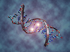
DNA methylation is a modification of a DNA molecule without changing the nucleotide sequence of DNA itself, which can be considered as part of the epigenetic component of the genome [1] [2] [3] .
DNA methylation involves the attachment of a methyl group to cytosine as part of a CpG dinucleotide at position C5 of the cytosine ring. Methylation in the promoter region of a gene, as a rule, leads to suppression of the corresponding gene. Methylated cytosine can then be oxidized by special enzymes, which ultimately leads to its demethylation back to cytosine [4] .
DNA methylation is considered mainly inherent in eukaryotes . In humans, about 1% of genomic DNA is methylated. In adult somatic cells, DNA methylation usually occurs in CpG dinucleotides ; DNA methylation outside of CpG dinucleotides is found in embryonic stem cells . [5] [6]
Moreover, DNA methylation outside of CpG dinucleotides is a hallmark of the epigenome of pluripotent stem cells, and a decrease in non-CG methylation is associated with impaired ability to differentiate into endoderm cell lines [7]
In plants, cytosine methylation occurs both symmetrically on both chains (on CpG or CpNpG), and asymmetrically on only one of the two chains (on CpNpNp, where N denotes any nucleotide).
Mammalian DNA methylation
About 60–70% of all CpG dinucleotides in mammals are methylated. Unmethylated CpG dinucleotides are grouped in the so-called. " CpG islands " that are present in the 5'- regulatory regions of many genes . Various diseases, such as cancer , are accompanied by initial abnormal DNA hypomethylation and subsequent hypermethylation of CpG islands in the promoter regions of the genes, which leads to a stable repression of transcription. Repression of transcription in this case is mediated by proteins that are capable of binding to methylated CpG dinucleotides. These proteins, called methylcytosine-binding proteins , attract histone deacetylase (HDAC) and other factors involved in chromatin remodeling. The formed complex can modify histones, forming a condensed transcriptionally inactive heterochromatin structure. The effect of DNA methylation on chromatin structure is of great importance for the development and functioning of a living organism. In particular, the absence of methylcytosine binding protein 2 (MeCP2) due to, for example, mutations in the corresponding gene, leads to the development of Rett syndrome in humans; inactivation of methylcytosine-binding domain protein 2 (Methyl-CpG binding domain protein 2 - MBD2), which is involved in the repression of transcription of hypermethylated genes, was noted in oncological diseases.
Human DNA Methylation
In humans, three enzymes responsible for DNA methylation are called DNA methyltransferases 1, 3a and 3b (DNMT1, DNMT3a, DNMT3b), respectively. It is assumed that DNMT3a and DNMT3b are de novo methyltransferases that carry out the formation of DNA methylation patterns in the early stages of development, as well as its changes during cell differentiation. There is a hypothesis that de novo DNA methylation is caused, in particular, by interfering RNAs using RNA-dependent DNA methylation, a process that arose during evolution to repress mobile genome elements. [8] DNMT1 is a DNA methyltransferase that maintains the methylated state of DNA by attaching methyl groups to one of the DNA strands at the points where the other strand complementary to it is methylated. It is assumed that the role of DNMT1 inhibitors that regulate DNA methylation is played by non-polyadenylated long non-coding RNAs (lncRNA) [9] The DNMT3L protein is homologous to other DNMT proteins, but does not have catalytic activity. Instead, DNMT3L supports de novo methyltransferases, promoting the binding of these enzymes to DNA and stimulating their activity.
Important stages in the development of malignant neoplasms are preliminary DNA hypomethylation [10] and subsequent inactivation of tumor suppressor genes [11] . In the case when inactivation was due to methylation of the promoter region of the gene, experiments were performed to resume expression by inhibiting DNMT. 5-aza-2'-deoxycytidine ( decitabine ) is a nucleoside analog that inhibits DNMT methyltransferases. The mechanism of action of the drug is based on covalent binding of the enzyme in complex with DNA, which makes it impossible for the enzyme to fulfill its function and leads to the degradation of methyltransferase. However, in order for decitabine to be active, it must be integrated into the genome of the cell, but this, in turn, can cause mutations in daughter cells if the cell does not die and continues to divide. In addition, decitabine is toxic to bone marrow, which narrows the scope of its therapeutic use. These limitations have led to an intensive search for therapeutic modalities based on the use of “antisense” RNAs that counteract DNMT through degradation of its mRNA and, therefore, block translation . The ability to selectively demethylate a gene and thus alter its expression is made possible by the discovery of the so-called extracoding RNA, which is able to bind to DNMT1, blocking its ability to methylate a specific gene [12] . Nevertheless, the question remains whether the inhibition of DNMT1 function is a sufficient condition for increasing the expression of suppressor genes, which are negatively regulated by DNA methylation.
Analysis of personal transcriptomes and human methylomas showed that only less than 20% of genes correlate between methylation and gene expression [13]
Young et al. have developed an effective method for the selective targeted demethylation of specific CpGs in human cells using the TALE domain (transcription activator-like effector) and the TET1 hydroxylase catalytic domain, catalyzing the conversion of 5-methylcytosine to 5-hydroxymethylcytosine, by molecular engineering [14] . Using this combined TALE-TET1 molecule, they showed that demethylation of certain CpG promoters can significantly increase the expression of the corresponding human genes. The development of an enzyme based on an inactivated d Cas9 molecule (incapable of cutting DNA) will greatly simplify the creation of selective demethylases. This will allow directing demethylase to the hypermethylated gene promoters selected for activation using the RNA guide. [15]
A medium has been developed that causes DNA hypomethylation in cells in vitro. This medium, called 2i, contains two low molecular weight inhibitors, one of which inhibits the ERK1 / 2 signaling pathway, and the other Gsk3β . It is widely used for reprogramming and maintaining the pluripotent state of cells [16] [17] .
DNA methylation changes during aging
Back in the 1970s, in the works of Vanyushin B.F. and a number of other employees of the biology faculty of Moscow State University , a relationship was found between the stages of individual development and aging of animals and humans with changes in such an enzymatic modification as DNA methylation [18] . For a series of works "DNA methylation - epigenetic control of the genetic functions of the body" Vanyushin B.F. was awarded the prize named after A.N. Belozersky .
It is now well known that the genomic DNA methylation landscape varies with age [19] [20] [21] [22] [23] . This process, called “epigenetic drift” [24] [25] , is closely associated with chronological age and, at the same time, according to some authors, it is not a marker of replicative cell aging , as it is also found in “non-aging” stem cells [26 ] [27] . For replicative cell aging, slightly different epigenetic biomarkers were found that are also based on changes in DNA methylation in certain places of the genome [28] Determination of epigenetic aging by DNA methylation of ITGA2B, ASPA, and PDE4C genes allows determining the biological age of a person with an average absolute deviation from the chronological age of not more than 5 years [29] . This accuracy is higher than age predictions based on telomere length.
During the aging process of the human body, the functional potential of hematopoietic stem cells (HSCs) is significantly reduced, which contributes to the development of hematopoietic pathophysiology in elderly people [30] . This age-related decrease in the number of HSCs and their ability to proliferate , as it turned out, depends to a greater extent not on telomere length, but on hypermethylation of DNA genes regulated by the Polycomb Repressor Complex 2 proteins [31] .
According to Chinese researchers [32] , DNA methylation can contribute to healthy human aging by regulating genes susceptible to age-related diseases. For example, in Alzheimer's disease , the caspase 3 CASP3 gene is actively expressed, which is involved in the cleavage of the precursor protein of beta-amyloid 4A, which leads to neuronal death, while in centenarians, the CASP3 gene is blocked by hypermethylation of the site near the transcription initiation site of this gene. Another example: the IL1R2 interleukin receptor gene has low expression in the case of atherosclerosis , while in long-livers its activity is not reduced due to the hypomethylated region near the site of its transcription initiation [32] [33] .
See also: Biological clock (aging) , as well as a popular science review in Russian [34] and a detailed review in English [35]
The role of methylation in oncogenesis
Epigenetic age, as it turned out, affects the likelihood of getting cancer. An algorithm has been developed to calculate epigenetic age from blood DNA methylation data. The algorithm is based on 71 DNA methylation markers, which may vary depending on the human environment, exercise, and diet. A study using this algorithm of a collection of blood samples collected over 15 years showed that those with an epigenetic age about one year older than their chronological age have a 6% greater risk of developing cancer for three years, and those who are about 2 2 years older than their chronological age have a 17% increased risk of dying from cancer within five years [36] [37] .
A comparison of the genotype data of people predisposed to oncological diseases with the methylation profile of their DNA suggests that in about 20% of cases of inherited cancer there is a relationship between the methylation level of certain loci and gene polymorphisms associated with cancer risk. In particular, a highly significant correlation was observed between the level of CpG methylation and the expression of key cancer genes such as MYC, TERT, and TP63 [38] .
About one third of all solid tumors are associated with a mutation of the KRAS gene or mutations in pathways associated with KRAS. KRAS turns off the TET1 enzyme, which promotes gene inactivation by methylation. TET1 catalyzes the initial stage of active iron- and alpha-ketoglutarate-dependent DNA demethylation in mammals - the conversion of 5-methylcytosine (5-MC) to 5-hydroxymethylcytosine (5-HMC) by oxidation of 5-MC. Adding TET1 to these mutant cells activates tumor suppressor genes, which, as it turns out, is enough to reduce abnormal proliferation. However, it is sufficient to inactivate TET1 to make these cells malignant again, even without KRAS [39] .
An important biomarker of oncogenesis is the hypermethylation of CpG islands within the promoter of the ZNF154 gene ( zinc-finger protein 154) [40] [41] . Hypermethylation of ZNF154 was observed in the vast majority of tumor cells, but was absent in normal cells [42] . What function in the body does the ZNF154 gene is not yet clear. At the same time, hypomethylation is usually observed in two genomic regions associated with Casp8 ( caspase -8) and VHL (suppressor of Hippel-Lindau tumors) [42] .
Insect DNA Methylation
The DNA methylation level in the favorite object of the geneticists Drosophila melanogaster is very low, which interfered with its study using bisulfite sequencing [43] . Takayama et al. [44] developed a highly sensitive method that revealed that the methylation profile of the DNA genome sequences of a fly is very different from the methylation profile of the human, animal, or plant genome. Drosophila genome methylation is concentrated in certain short base sequences (of 5 pairs of nucleotides) that are rich in CA and CT, but are depleted in guanine. In addition, it turned out to be independent of the activity of DNMT2. Further study of DNA methylation in Drosophila will help identify age-related changes in the epigenome.
DNA methylation in plants
Recently, there has been a significant breakthrough in understanding the process of DNA methylation in plants, especially in Arabidopsis thaliana . The main DNA methyltransferases in A. thaliana are Met1, Cmt3, and Drm2, which at the amino acid sequence level are similar to mammalian DNA methyltransferases described above. Drm2 is thought to be involved in both de-novo DNA methylation and maintenance of methylation at later stages of development. Cmt3 and Met1 mainly fulfill the function of maintaining DNA methylation. [45] Other DNA methyltransferases are also present in plants, but their function has not yet been elucidated (See [1] ). It is believed that the specificity of methyltransferase during DNA methylation is modulated by RNA. Specific RNA transcripts are transcribed from certain parts of the matrix - genomic DNA. These RNA transcripts can form double-stranded RNA molecules. Double-stranded RNAs, through regulatory signaling pathways associated with either small interfering RNAs (siRNAs) or microRNAs (miRNAs), determine the location of DNA methyltransferases in regions of specific nucleotide sequences in the genome [46] .
See also
- Gene imprinting
- Reprogramming
- DNA methyl transferase
- 5-hydroxymethylcytosine
- Bisulfite sequencing
Notes
- ↑ Bethany A. Buck-Koehntop and Pierre-Antoine Defossez (2013) On how mammalian transcription factors recognize methylated DNA. 8 (2), 131-137 https://dx.doi.org/10.4161/epi.23632
- ↑ Seisenberger S, Peat JR, Hore TA, Santos F, Dean W, Reik W. (2013) Reprogramming DNA methylation in the mammalian life cycle: building and breaking epigenetic barriers. Phil. Trans. R. Soc. B 368, 20110330. doi: 10.1098 / rstb.2011.0330
- ↑ Kelsey G, Feil R. (2013) New insights into establishment and maintenance of DNA methylation imprints in mammals. Phil. Trans. R. Soc. B 368, 20110336. doi: 10.1098 / rstb.2011.0336
- ↑ Rahul M. Kohli & Yi Zhang (2013) TET enzymes, TDG and the dynamics of DNA demethylation. Nature 502 (7472), 472-479 doi: 10.1038 / nature12750
- ↑ Dodge, Jonathan E .; Bernard H. Ramsahoyeb, Z. Galen Woa, Masaki Okanoa, En Li. De novo methylation of MMLV provirus in embryonic stem cells: CpG versus non-CpG methylation (Eng.) // ScienceDirect : journal. - 2002. - May.
- ↑ Haines, Thomas R. Allele-Specific Non-CpG Methylation of the Nf1 Gene during Early Mouse Development (Eng.) // ScienceDirect : journal. - 2001. - December.
- ↑ Lee M. Butcher, Mitsuteru Ito, Minodora Brimpari, Tiffany J. Morris, Filipa AC Soares, Lars Ährlund-Richter, Nessa Carey, Ludovic Vallier, Anne C. Ferguson-Smith, Stephan Beck. (2016). Non-CG DNA methylation is a biomarker for assessing endodermal differentiation capacity in pluripotent stem cells. Nature Communications 7, Article number: 10458 DOI : 10.1038 / ncomms10458
- ↑ Galitsky V.A. A hypothesis on the mechanism of small RNA initiation of de novo DNA methylation and allelic exclusion (rus.) // Cytology. - 2008 .-- T. 50 (4) . - S. 277-286 .
- ↑ Fan Lai and Ramin Shiekhattar (January 2014). Where long noncoding RNAs meet DNA methylation . Cell Research, doi: 10.1038 / cr.2014.13
- ↑ Elias Daura-Oller, Maria Cabre, Miguel A Montero, Jose L Paternain, and Antoni Romeu (2009) "Specific gene hypomethylation and cancer: New insights into coding region feature trends." Bioinformation. 2009; 3 (8): 340—343.PMID PMC2720671
- ↑ Stepanenko, AA, & Kavsan, VM (2012) Immortalization and malignant transformation of Eukaryotic cells. Cytology and Genetics, 46 (2), 96-129
- ↑ Di Ruscio, A., Ebralidze, AK, Benoukraf, T. Et al. & Tenen, DG (2013) DNMT1-interacting RNAs block gene-specific DNA methylation . Nature; DOI: 10.1038 / nature12598
- ↑ Rubina Tabassum, Ambily Sivadas, Vartika Agrawal, Haozheng Tian, Dalia Arafat and Greg Gibson (2015). Omic personality: implications of stable transcript and methylation profiles for personalized medicine . Genome Medicine 2015, 7:88 DOI : 10.1186 / s13073-015-0209-4
- ↑ Maeder, ML, Angstman, JF, Richardson, ME, et al. & Joung, JK (2013). Targeted DNA demethylation and activation of endogenous genes using programmable TALE-TET1 fusion proteins. Nature biotechnology. doi: 10.1038 / nbt.2726
- ↑ Xu, X., Tao, Y., Gao, X., Zhang, L., Li, X., Zou, W., ... & Hu, R. (2016). A CRISPR-based approach for targeted DNA demethylation . Cell Discovery, 2., Article number: 16009 (2016) DOI : 10.1038 / celldisc.2016.9
- ↑ Gabriella Ficz, Timothy A. Hore, Fátima Santos, et al. & Wolf Reik (2013) FGF Signaling Inhibition in ESCs Drives Rapid Genome-wide Demethylation to the Epigenetic Ground State of Pluripotency . Cell Stem Cell, 13(3), 351—359 https://dx.doi.org/10.1016/j.stem.2013.06.004
- ↑ Hakan Bagci, Amanda G. Fisher (2013) DNA Demethylation in Pluripotency and Reprogramming: The Role of Tet Proteins and Cell Division. Cell Stem Cell, 13(3), 265—269 doi: 10.1016/j.stem.2013.08.005
- ↑ Ванюшин Б., Бердышев Г. (1977). Молекулярно-генетические механизмы старения . Изд-во Медицина. Moscow. Букинистическое издание.
- ↑ Johansson Å, Enroth S, Gyllensten U (2013) Continuous Aging of the Human DNA Methylome Throughout the Human Lifespan. PLoS ONE 8(6): e67378. doi:10.1371/journal.pone.0067378
- ↑ Hannum, G., Guinney, J., Zhao, L., et al. & Zhang, K. (2013). Genome-wide methylation profiles reveal quantitative views of human aging rates. Molecular cell, 49(2), 359—367.
- ↑ Heyn H, Li N, Ferreira HJ, Moran S, Pisano DG, et al. (2012) Distinct DNA methylomes of newborns and centenarians. Proc Natl Acad Sci USA 109: 10522-10527. doi:10.1073/pnas.1120658109
- ↑ Pogribny, IP, & Vanyushin, BF (2010). Age-related genomic hypomethylation. In Epigenetics of Aging (pp. 11-27). Springer New York. DOI: 10.1007/978-1-4419-0639-7_2
- ↑ Florath, I., Butterbach, K., Müller, H., Bewerunge-Hudler, M., et al. & Baca, V. (2013). Cross-sectional and longitudinal changes in DNA methylation with age: An epigenome-wide analysis revealing over novel age-associated CpG sites. Human molecular genetics, doi: 10.1093/hmg/ddt531
- ↑ Teschendorff, AE, West, J., & Beck, S. (2013). Age-associated epigenetic drift: implications, and a case of epigenetic thrift?. Hum. Mol. Genet. 22 (R1):R7-R15 doi: 10.1093/hmg/ddt375
- ↑ West, J., Widschwendter, M., & Teschendorff, AE (2013). Distinctive topology of age-associated epigenetic drift in the human interactome. PNAS, 110(35), 14138-14143. doi:10.1073/pnas.1307242110
- ↑ Steve Horvath (2013) DNA methylation age of human tissues and cell types . Genome Biology, 14(10):R115 doi:10.1186/gb-2013-14-10-r115
- ↑ Bocklandt S, Lin W, Sehl ME, Sánchez FJ, Sinsheimer JS, et al. (2011) Epigenetic Predictor of Age . PLoS ONE 6(6): e14821. doi:10.1371/journal.pone.0014821
- ↑ Koch, CM, & Wagner, W. (2013). Epigenetic Biomarker to Determine Replicative Senescence of Cultured Cells. In Biological Aging. Сер.: Methods in Molecular Biology, Vol. 1048, (pp. 309—321). Humana Press. DOI: 10.1007/978-1-62703-556-9_20
- ↑ Carola Ingrid Weidner, Qiong Lin, Carmen Maike Koch et al. and Wolfgang Wagner (February 2014). Aging of blood can be tracked by DNA methylation changes at just three CpG sites . Genome Biology, 15:R24 doi:10.1186/gb-2014-15-2-r24
- ↑ Myung Geun Shin (2014). Aging and impaired hematopoiesis. J Korean Med Assoc.; 57(4), 334-340. https://dx.doi.org/10.5124/jkma.2014.57.4.334
- ↑ Beerman, I., Bock, C., Garrison, BS, Smith, ZD, Gu, H., Meissner, A., & Rossi, DJ (2013). Proliferation-dependent alterations of the DNA methylation landscape underlie hematopoietic stem cell aging. Cell Stem Cell, 12(4), 413-425 DOI : 10.1016/j.stem.2013.01.017
- ↑ 1 2 Xiao FH, He YH, Li QG, Wu H, Luo LH, Kong QP (2015). A Genome-Wide Scan Reveals Important Roles of DNA Methylation in Human Longevity by Regulating Age-Related Disease Genes . PLoS ONE 10(3): e0120388. DOI : 10.1371/journal.pone.0120388
- ↑ Jones, MJ, Goodman, SJ and Kobor, MS (2015), DNA methylation and healthy human aging . Aging Cell. DOI : 10.1111/acel.12349
- ↑ Определение биологического возраста человека по метилированию ДНК
- ↑ Horvath, S., & Raj, K. (2018). DNA methylation-based biomarkers and the epigenetic clock theory of aging. Nature Reviews Genetics, DOI : 10.1038 / s41576-018-0004-3 PMID 29643443
- ↑ Epigenetic-Chronological Age Mismatch Warns of Cancer . Genetic Engineering & Biotechnology News. Feb 18, 2016
- ↑ Zheng, Y., Joyce, BT, Colicino, E., Liu, L., Zhang, W., Dai, Q., ... & Jafari, N. (2016). Blood Epigenetic Age may Predict Cancer Incidence and Mortality. EBioMedicine. DOI : 10.1016 / j.ebiom.2016.02.00.008
- ↑ Heyn H, Sayols S, Moutinho C, et al. & Esteller M. (2014) Linkage of DNA Methylation Quantitative Trait Loci to Human Cancer Risk. Cell Reports, https://dx.doi.org/10.1016/j.celrep.2014.03.01.016
- ↑ Bo-Kuan Wu, Charles Brenner (2014). Suppression of TET1-Dependent DNA Demethylation Is Essential for KRAS-Mediated Transformation. Cell Reports, DOI: https://dx.doi.org/10.1016/j.celrep.2014.10.063
- ↑ Gennady Margolin, Hanna M. Petrykowska, Nader Jameel, Daphne W. Bell, Alice C. Young, Laura Elnitski (2016). Robust Detection of DNA Hypermethylation of ZNF154 as a Pan-Cancer Locus with in Silico Modeling for Blood-Based Diagnostic Development. The Journal of Molecular Diagnostics. DOI: https://dx.doi.org/10.1016/j.jmoldx.2015.11.004
- ↑ Researchers identify striking genomic signature shared by five types of cancer
- ↑ 1 2 Sánchez-Vega, F., Gotea, V., Petrykowska, HM, Margolin, G., Krivak, TC, DeLoia, JA, ... & Elnitski, L. (2013). Recurrent patterns of DNA methylation in the ZNF154, CASP8, and VHL promoters across a wide spectrum of human solid epithelial tumors and cancer cell lines. Epigenetics, 8 (12), 1355-1372. DOI : 10.4161 / epi.26701
- ↑ Capuano F., Mülleder M., Kok RM, Blom HJ, Ralser M. Cytosine DNA methylation is found in Drosophila melanogaster but absent in Saccharomyces cerevisiae , Schizosaccharomyces pombe and other yeast species (Eng.) // Analytical Chemistry: journal. - 2014 .-- Vol. 86 , no. 8 . - P. 3697-3702 . - DOI : 10.1021 / ac500447w . - PMID 24640988 .
- ↑ S. Takayama, J. Dhahbi, A. Roberts, G. Mao, S.-J. Heo, L. Pachter, DIK Martin, D. Boffelli (2014). Genome methylation in D. melanogaster is found at specific short motifs and is independent of DNMT2 activity. Genome Research, DOI : 10.1101 / gr. 162412.113
- ↑ Cao, Xiaofeng; Jacobsen, Steven E. Locus-specific control of asymmetric and CpNpG methylation by the DRM and CMT3 methyltransferase genes (English) // Proceedings of the National Academy of Sciences : journal. - Jul. - 2003.
- ↑ Aufsatz, Werner; M. Florian Mette, Johannes van der Winden, Antonius JM Matzke, Marjori Matzke. RNA-directed DNA methylation in Arabidopsis (neopr.) // Proceedings of the National Academy of Sciences . - Dec. - 2002.
Literature
- Machado, ACD, Zhou, T., Rao, S., Goel, P., Rastogi, C., Lazarovici, A., ... & Rohs, R. (2014). Evolving insights on how cytosine methylation affects protein – DNA binding . Briefings in functional genomics, elu040. DOI : 10.1093 / bfgp / elu040
- Smith, ZD & Meissner, A. (2013). DNA methylation: roles in mammalian development. Nat. Rev. Genet. 14, 204–220 DOI : 10.1038 / nrg3354 PMID 23400093
- Wu, H., & Zhang, Y. (2014). Reversing DNA Methylation: Mechanisms, Genomics, and Biological Functions. Cell, 156 (1), 45-68. https://dx.doi.org/10.1016/j.cell.2013.12.01.01
- Heather J. Lee, Timothy A. Hore, Wolf Reikemai (June 2014). Reprogramming the Methylome: Erasing Memory and Creating Diversity . Cell Stem Cell, 14 (6), 710-719, https://dx.doi.org/10.1016/j.stem.2014.05.008
- Paul M. Lizardi, Qin Yan, and Narendra Wajapeyee (2012) Analysis of DNA Methylation in Mammalian Cells. In: Molecular Cloning: A Laboratory Manual (Fourth Edition), VOLUME 2, CHAPTER 12, 893–942, ISBN 978-1-936113-42-2 A detailed description of the methods.
- Jiwook Shim, Gwendolyn I. Humphreys, Bala Murali Venkatesan, et al & Rashid Bashir (2013). Detection and Quantification of Methylation in DNA using Solid-State Nanopores . Scientific Reports, 3, Article number: 1389, DOI: 10.1038 / srep01389 In the preface, an overview of the methods.
- Jaroslav Fulneček * and Aleš Kovařík (2014). How to interpret Methylation Sensitive Amplified Polymorphism (MSAP) profiles? BMC Genetics, 15: 2 DOI : 10.1186 / 1471-2156-15-2 According to the authors, a reliable, inexpensive and relatively simple method for studying DNA methylation.
- Szulwach, KE and Jin, P. (2014), Integrating DNA methylation dynamics into a framework for understanding epigenetic codes . Bioessays 36: 107-117. DOI : 10.1002 / bies.201300090
- Nathan R. Rose, Robert J. Klose, (2014). Understanding the relationship between DNA methylation and histone lysine methylation . Biochimica et Biophysica Acta (BBA) - Gene Regulatory Mechanisms DOI : 10.1016 / j.bbagrm.2014.02.00.007
- ANNA GOOD WOMAN (2018). “We have penetrated the secrets of DNA to understand how fast people are aging,” geneticist Steve Horvath . “KP” [part 1], 03/26/2018 2:41 a.m.
- ANNA GOOD WOMAN (2018). The geneticist who created the DNA clock for aging, Steve Horvath: everyday stress and red meat do not steal our youth . “KP” [part 2]
- Dmitry Dzhagarov (2016). DNA methylation and some other epigenetic mechanisms that regulate aging . Scientific Expert Group RLE
- Dmitry Dzhagarov (2016). CRISPR / Cas9 and epigenetics . "KhiZh", 2016 №12
- Levine, ME, Lu, AT, Quach, A., Chen, B., Assimes, TL, Bandinelli, S., ... & Whitsel, EA (2018). An epigenetic biomarker of aging for lifespan and healthspan. bioRxiv, 276162. DOI : 10.1101 / 276162
- Declerck, K., & Berghe, WV (2018). Back to the future: epigenetic clock plasticity towards healthy aging . Mechanisms of ageing and development. DOI : 10.1016 / j.mad.2018.01.002 A detailed review of epigenetic clocks built on DNA methylation
- Han, Y., Eipel, M., Franzen, J., Sakk, V., Dethmers-Ausema, B., Yndriago, L., ... & Wagner, W. (2018). Epigenetic age-predictor for mice based on three CpG sites . eLife, 7, e37462. DOI : 10.7554 / eLife.37462
- Aquino, EM, Benton, MC, Haupt, LM, Sutherland, HG, Griffiths, LR, & Lea, RA (2018). Current understanding of DNA methylation and age-related disease. OBM Genetics, 2 (2). DOI : 10.21926 / obm.genet.1802016 Modern ideas about DNA methylation are very clearly outlined. Especially in Fig. 2
- Field AE, Robertson NA, et al., & Adams PD (2018). DNA Methylation Clocks in Aging: Categories, Causes, and Consequences . Molecular Cell. 71 (6), 882-895, DOI: https://doi.org/10.1016/j.molcel.2018.08.008