Actinomycetes ( obsolete. Radiant fungi ) (from actinomycetes + mycetes) - the order of bacteria that have the ability to form at some stages of development of branching mycelium , which manifests itself in optimal conditions for their existence. Some researchers, emphasizing the bacterial nature of actinomycetes, call their analogue of mushroom mycelium thin threads ; their diameter is 0.4-1.5 microns. Actinomycetes have an acid-resistant type of cell wall , which is Gram stained as gram-positive , but closer in structure to gram-negative. They are characterized by a high (60–75%) content of H C pairs in DNA .
| Actinomycetes | |||||||||
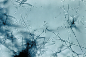 Streptomyces sp. | |||||||||
| Scientific classification | |||||||||
|---|---|---|---|---|---|---|---|---|---|
| |||||||||
| International scientific name | |||||||||
Actinomycetales Buchanan 1917 | |||||||||
The most common in the soil : representatives of almost all genera of actinomycetes are found in it. Actinomycetes usually make up a quarter of the bacteria that grow on traditional media when their diluted soil suspensions are seeded and 5-15% of the prokaryotic biomass determined by luminescence microscopy . Their environmental role is most often the decomposition of complex, resistant substrates; presumably they are involved in the synthesis and decomposition of humic substances . They can act as symbionts of invertebrates and higher plants .
Content
- 1 Building
- 2 Life cycle
- 2.1 Spore formation
- 2.2 Germination of spores
- 3 Mobility
- 4 Environmental functions
- 5 Attitude to environmental factors
- 6 Biochemical features
- 7 Actinomycete genera groups according to the 9th edition of the Bergee determinant
- 7.1 Nocardioform actinomycetes
- 7.2 Births with multi-nest sporangia
- 7.3 Actinoplanes
- 7.4 Streptomycetes and related birth
- 7.5 Maduroomycetes
- 7.6 Thermal monospores and related birth
- 7.7 Genus Termoactynomyces
- 8 History of study
- 9 notes
- 10 Literature
Building
Indirect data suggest apical growth in actinomycetes.
The differentiation of mycelium is a process of complication in the development of an actinomycete colony. First of all, it manifests itself in the division into primary (substrate) and secondary (air) mycelium. The air is thicker, it is hydrophobic , contains more DNA and enzymes, on the surface of its cells there are various structures (rod-shaped, fibrils).
Mycelium with rare septa, almost valuable in spore-forming, with frequent septa (septa) in forms for which the mycelium disintegrates and are close to them. Vegetative cells of most forms are divided by transverse septa, Geodermatophilus and Dermatophilus in mutually perpendicular directions, some actinomycetes contain cells with septa that extend in completely different directions (sporangia Micromonospora , Frankia vesicles). Branching occurs by the mechanism of budding.
Often, differentiation is manifested in the formation of amicellar structures:
- coremia - a close interweaving of fused hyphae glued together by mucus with iron oxides;
- cell aggregates;
- crystals of secondary metabolites;
- "Sulfur granules" ;
- sclerotia - thickened hyphae with vacuoles filled with lipids can grow as a spore;
- vesicles are encapsulated nitrogen-fixing formations in Frankia .
In the process of aging, the cytoplasm of cells acquires an uneven electron density, ribosomes cease to be distinguished in it, the nucleoid boundary spreads, the cell wall becomes thin and loose, and a microcapsule forms. During autolysis in the cytoplasm, extensive bright areas are formed, the nucleoid decays, holes form in the cell wall, the cell is filled with membrane structures that are destroyed by the latter.
Life Cycle
Nocardioform actinomycetes rarely form spores and multiply mainly by fragments of rapidly decaying mycelium. Actinomycetes with prolonged mycelial stages differ in the type of spore formation.
Spore formation
According to the number of spores, actinomycetes are divided into mono- (for example, Saccaromonospora , Micromonospora ), oligo- ( Actinomadura ) and polyspore ( Streptomyces ), highlighting especially those that form sporangia. Spore formation is predominantly exogenous ( Thermoactinomyces forms true endospores, however, at present, this genus, on the basis of chemotaxonomic and genetic characters, despite the pronounced mycelial stage, is prone to be referred to as bacilli), less often pseudo-endogenous ( Planomonospora , Dactylosporangium ).
Streptomyces and sporulating Actinomyces spores form in two stages:
- The apical section of the air hyphae is separated by a septum, the nucleoid is extended.
- Almost simultaneously, the cell divides into septa, the nucleoid divides in the same places, the cell wall becomes 2 times thicker, the spores are rounded and their wall becomes 7 times thicker than the wall of the hyphae.
In oligospore septa, they are laid basipitally. Monsporovy can be formed by the mechanism of budding.
Spore formation is caused by the so-called factor A (C 13 H 22 O 4 ).
Germination of spores
Germination occurs in the following stages:
- Inactive spore hydrophobic, heat-resistant, does not show respiratory activity
- Wettable activated spore shows enzyme activity, respiration begins
- Spore swells, RNA synthesis begins
- Yield 1-3 (less often 4) growth tubes, DNA synthesis begins. This stage is irreversible, the other three are reversible.
Druze form a cluster of interwoven filaments with bulbous thickenings at the ends.
Mobility
They can be mobile at the stage of spores ( actinoplanes , Geodermatophilus and Dermatophilus ), sometimes parts of the mycelium ( Erskovia ) are mobile.
Environmental Functions
Actinomycetes (especially the genus Micromonospora ) are found in water bodies and their bottom sediments, but the question of whether they are their permanent inhabitants or brought from the soil is not resolved, their role in these habitats is also unknown.
Soils are the natural substrate from which actinomycetes stand out in the greatest variety. However, most of the biomass of actinomycetes is represented by spores, which give colonies when counting populations in the soil by sowing, only 1-4% of the biomass is mycelium [1] . It is found in microzones with a high content of organic matter.
Actinomycetes dominate in the late stages of microbial succession , when conditions are created for the use of hard-to-reach substrates. Activation of actinomycete microflora occurs when starch , chitin , oil products, etc. are introduced into the soil. At the same time, due to slow growth, actinomycetes are not able to compete with non-mycelial bacteria for readily available substances. It is possible that secondary metabolites (especially melanoid pigments ) play some role in the formation of humus .
Actinomycetes play a price-forming role in places of primary soil formation, being under these conditions in association with algae. Under laboratory conditions, these associations formed a lichen-like thallus ( actinolenic ).
Actinomycetes (the genus Streptomyces , Streptosporangium , Micromonospora , Actinomadura ) are permanent inhabitants of the intestines of earthworms , termites and many other invertebrates . Destroying cellulose and other biopolymers, they are their symbionts . Representatives of the genus Frankia are capable of nitrogen fixation and the formation of nodules in non-leguminous plants ( sea buckthorn , alder , etc.). There are pathogenic forms that cause actinomycosis . In the human body they live in the oral cavity, in the intestines, in the respiratory tract, on the skin, in plaque, in carious teeth, on the tonsils.
Attitude to environmental factors
Most actinomycetes are aerobes ; facultative anaerobes are present only among actinomycetes with a short mycelial stage. There is a certain parallel with mushrooms , among which only non-mycelial yeast is also able to live in anaerobic conditions. A less efficient anaerobic type of metabolism is thought to be successful with a larger relative cell surface, which is achieved by fragmentation of the mycelium.
Actinomycetes are believed to be more resistant to drying than non-mycelial bacteria, which is why they dominate in desert soils. Laboratory storage of soil samples under conditions not conducive to the vegetative growth of prokaryotes increases the relative content of actinomycetes taken into account by the inoculation method. Especially for a long time, sclerotia formed by the genus Chainia can persist during drying. It was shown that at a w = 0.50, some spores germinate ( Streptomyces , Micromonospora ), but the formed mycelium does not branch. At a w = 0.86, spores of almost all actinomycetes germinate, in some mycelia it branches, microcolonies form, the optimum is reached at a w = 0.95.
Most often, neutrophils actinomycetes, however, some genera are acidophilic or alkalophilic. A characteristic property of actinomycetes is acid tolerance, due to which their share in the microbial complex of forest soils is relatively high. It was noted that on the acidic medium the vegetative stage is prolonged, on the alkaline, on the contrary, spore formation is accelerated.
Actinomycetes are not demanding on the content of organic carbon in the environment, many of them are able to grow on "hungry" agar-agar . Representatives of the genus Nocardia are able to carry out chemosynthesis by oxidizing hydrogen , methane and methanol . Heterotrophic fixation of CO 2 is widespread among actinomycetes.
Biochemical features
For actinomycetes, the presence of rare metabolic pathways and enzyme systems is noted. For example, they are characterized by the pathway of the breakdown of Entner-Dudorov glucose , polyphosphate hexokinase is found (instead of the usual hexokinase ), there are features in the synthesis of a number of amino acids; in the secondary metabolism, they are characterized by a shikimate pathway for the synthesis of aromatic compounds, the inclusion of whole carbon skeletons of glucose in secondary metabolites, for example, antibiotics.
A distinctive feature of actinomycetes is the ability to synthesize physiologically active substances, antibiotics, pigments, odorous compounds. It is they who form the specific smell of soil and sometimes water (substances geosmin , argosmin , mucidon , 2-methyl-isoborneol ). Actinomycetes are active producers of antibiotics , forming up to half known to science.
Actinomycete genera groups according to the 9th edition of the identifier Bergey
Nocardioform actinomycetes
Aerobic organisms having a mycelial stage in the development cycle. Mycelium can decay into elements, forming chains similar to sporangia. There is no real argument. This includes the genus Nocardia , Rhodococcus , which is able to use oil hydrocarbons, Promicromonospora , Actinobispora , Oerskovia , etc. The division into genera is based on the chemotype of the cell wall and other chemotaxonomic characters.
Genera with multi-nest sporangia
The resulting mycelium is divided into individual coccoid cells motile in Geodermatophilus and Dermatophilus and motionless in Frankia . Francia - nitrogen - fixing symbionts of alder and other non-leguminous plants, forming nodules on their roots. Habitat: soil, water and skin of mammals .
Actinoplanes
In the development cycle, they have a mobile stage and the stage of formation of a developed mycelium, divided by partitions. Saprotrophs and facultative parasites . Distributed in soil, forest litter , animal remains and water from natural sources, often developing on the pollen of plants caught in it. Divided into childbirth by types of sporangia:
- Mobile spores in sporangia ( Actinoplanes , Ampullariella , Dactylosporangium , Pilimelia )
- Fixed disputes
- Single ( Micromonospora )
- In Chains ( Catellatospora )
Cell wall type II (contains meso-DAPK and glycine ).
Streptomycetes and related births
They form a well-developed aerial mycelium, which does not decay into individual cells during development. Sporangia consist of straight or spiral-bound chains of motionless spores. They live in the soil and are characterized by strong antibiotic and chitin-degrading activity.
Type I cell wall (contains L-DAPK)
Maduroomycetes
Mycelium also does not decompose into individual cells. Disputes only on aerial mycelium in chains or sporangia, both mobile and not. The group is poorly studied and requires an audit. They form short chains of spores ( Actinomadura et al.), Sporangia with immobile ( Planomonospora ) or motile spores ( Streptosporangium ).
Types of cell walls II — IV. Madurosis is found in the hydrolysates of whole cells.
Thermomonospores and related births
Developed mycelium, spores are located singly, in chains or sporangi-like structures. Type III cell wall (meso-DAPK, no differentiating sugars).
Genus Termoactynomyces
Thermoactinomycetes form typical endospores and, according to this feature, as well as the structure of 16s rRNA, should be referred to as bacilli , however, they form a developed mycelium. Thermophiles capable of growing in the range of 40–48 degrees Celsius.
Study History
In 1874, F. Cohn in a sample from the human tear duct first discovered a filamentous bacterium, named after the doctor who took the Streptothrix foersteri sample. Since the generic name Strepothrix was already occupied by the fungus, the bacterium was later renamed Streptomyces foersteri . In 1877, the pathologist Bollinger and the botanist Harz examined the tumors (actinomycotic nodes) of cows and found their causative agent, which, due to the radiant arrangement of the strands, was called the radiant fungus ( Actinomyces ). This name soon became collective for several close births.
In 1884, the German physician James Israel received the first pure culture of actinomycete ( Actinomyces israelii ). In the future, many pathogenic forms were discovered (in 1888, the first representative of the genus Nocardia was isolated from the leg of a person suffering from Maduro disease ), in 1890-1892 Gospirini compiled a list of genera of actinomycetes.
In 1912-1916, the first descriptions of non-pathogenic actinomycetes isolated from ordinary natural substrates began to appear. During this period, S. A. Waxmann , Krainsky, Rudolf Liske contributed to the development of actinomycetology.
A new stage in the development of science began in 1939, when Krasilnikov received in his native form the antibiotic mycetin secreted by streptomycetes. In 1945, Waxman, Schatz and Boogie isolated streptomycin . Great attention was paid to actinomycetes, however, the applied aspects of actinomycetology related to the production and use of antibiotics mainly developed. Nevertheless, during this period, information was also obtained on the ecology, biochemistry, structure, development cycles, which in turn allowed the development of principles for the classification of actinomycetes.
From the 1980s and 1990s, attention shifted to the study of the ecological functions of actinomycetes, their relationships in natural conditions with animals, plants, and microorganisms. A systematic review is underway to obtain data on the genome of actinomycetes.
Notes
- ↑ Zvyagintsev D.G. Soil and microorganisms. - M .: 1987
Literature
- Zenova G.M. Soil Actinomycetes. - M .: Publishing house of Moscow State University, 1992. - ISBN 5-211-02902-X .
- The determinant of the bacteria Bergee. In 2 vols. T. 2: Trans. from English / Ed. J. Hole, N. Krieg, P. Snit, J. Staley, S. Williams. - M .: Mir, 1997 .-- 368 p., Ill. - ISBN 5-03-003112-X .