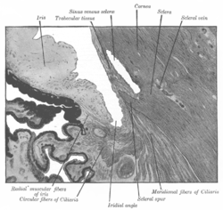
Trabecular network ( lat. Pectinatum anguli iridocornealis ) (JNA) is a reticular connective tissue that connects the ciliary edge of the iris with the edge of the posterior surface of the cornea and through which the aqueous humor of the anterior chamber of the eyeball is filtered into the Schlemm canal .
Formed from spongy fabric .
The second path of the outflow, uveoscleral (5-10%), helps the trabecular network to a small extent. The outflow along the uveoscleral pathway is accelerated by using certain medications for glaucoma, in particular, prostaglandins (for example, taflotan, xalatan, travatan).
Build
The trabecular network is divided into three parts with characteristically different ultrastructures:
- Internal uveal network - located closer to the angle of the anterior chamber of the eye, formed from thin heavy plates, oriented mainly radially.
- Corneoscleral network - contains a large amount of elastin , placed in the form of several layers of thin, flat, perforated sheets. It is considered a ciliary muscle tendon [1] .
- Juxtacanalicular network - adjacent directly to the channel Schlemm . This is a thin connective tissue band, covered with a single layer of epithelial cells. Chemical composition rich in glycosaminoglycans andglycoproteins .
Significance of glaucoma
Glaucoma occurs when intraocular pressure increases. May be caused due to increased formation of aqueous humor or its slow suction. Trabecular meshwork removes most of the aqueous humor.
In glaucoma, moisture is filtered through the transition zone of the Descemet's membrane and through the trabecular meshwork in the area of the opened Schlemm's canal.
Notes
- ↑ Sampaolesi R, Sampaolesi JR, Zárate G (2009). “Ocular Embryology with Special Reference to Chamber Angle Development” (chapter 8). The Glaucomas - Pediatric Glaucomas (volume 1). Springer Berlin Heidelberg. pp. 61-69. ISBN 978-3-540-69146-4 . http://www.springerlink.com/content/rkp83w0822835376/