Loricefera [1] ( lat. Loricifera ) is a class of marine animals of the type Scalidophora or Cephalorhyncha [2] . Some taxonomists consider taxon in the rank of type . These are very small (less than 0.5 mm) interstitial marine animals. About 80 species are known [1] [3] .
| Loricefera | |||||||||||||||||
 | |||||||||||||||||
| Scientific classification | |||||||||||||||||
|---|---|---|---|---|---|---|---|---|---|---|---|---|---|---|---|---|---|
| |||||||||||||||||
| International Scientific Name | |||||||||||||||||
Loricifera Kristensen , 1983 | |||||||||||||||||
| Squads and families | |||||||||||||||||
| |||||||||||||||||
Content
Build
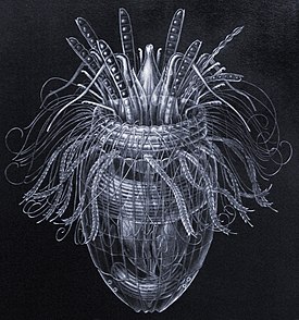
The body of loritsifer consists of almost 10 thousand cells , but in size they are equal to large infusoria (from 80 to 400 microns ) [3] . The body is divided into trunk and torso, between which is the chest region. At the front end of the trunk there is a retractable oral cone. The trunk can also be pulled into the front end of the shell. The screwing in of the trunk is provided by 30 muscles, which are combined into two groups. On the trunk is located up to 300 complex skalid, which probably perform sensory and locomotor functions. Some Scaleids have internal muscles [1] .
On top of the epidermis lies the chitinous cuticle, and underneath it underlay individual muscle cells . The most developed cuticle is located on the surface of , the trunk shell. Externally, Lorica resembles the shell of rotifers , but in loricifer it is extracellular, and in rotifers it is intracellular. Lorica consists of either 6 or 22 two long, parallel to the cuticle plates. In the area of lorica, the cuticle consists of a sclerotized epicuticle, intracuticle, and procutikula. At the junction of the plates, the cuticle remains flexible and non-sclerotized. The epidermis is represented by a single-layer epithelium , separated from the connective tissue by the basement membrane . Under the basement membrane are the muscles of the body. They are represented by longitudinal fibers in combination with diagonal, spinal-abdominal or ring fibers [4] .
The digestive system includes the frontal gut lined with the cuticle, the endodermal midgut and the cuticle of the posterior intestine. The mouth is located at the end of the oral cone and leads into the oral cavity, followed by a bulbous pharynx, consisting of epithelial-muscle cells and having a three-beam view of the cross section. The pharynx passes into a short cuticularized esophagus leading to the middle intestine. Medium intestine, lined with microvillary suction ; it accounts for most of the intestinal tube. The posterior intestine is short and opens outwardly with the anus near the posterior end of the body. Judging by the structure of the pharynx and oral cone, loricifera are carnivorous animals that suck the juices from the victim [5] .
The nervous system lies in the thickness of the epidermis. The brain consists of three rings and fills most of the trunk. The ganglionic forebrain (anterior ring) innervates the trunk and skalida. The midbrain is a fibrous neuropil without ganglia. The hindbrain consists of 10 ganglia, from which 10 longitudinal nerve cords depart, and two mid-abdominal cords form a double ganglionized nerve cords [5] .
Reproduction and development
Loricefera are semi-sexual, in some species even sexual dimorphism is expressed , which manifests itself in the structure and location of the skalid. Like priapulids , the excretory organs and genitals are combined into the urogenital system . Gonads are represented by paired sacks, which include the epithelium, which gives rise to gametes , and protonephridia . Gametes and urine enter the general urogenital tract, which opens with or near the anus. Seed receivers were found in one species, therefore fertilization , apparently, is internal [5] .
Features of embryonic development are unknown. A special type of larva emerges from the egg, resembling an adult, the Higgins larva . Its rear end is equipped with appendages called fingers. In some species, the fingers are wide, supplied with the muscles of the blade. Perhaps fingers are used for swimming. Other representatives on the tips of the thin fingers open glands, secreting a sticky secret that serves to attach to the substrate [5] .
Distribution and habitat
Loricefera are widely distributed in sediments of different types (sandy or silty) at different depths from the tropics to polar waters. The density of their populations is extremely low, and individual individuals are very firmly attached to the substrate particles, so it is very difficult to find them. Some species were found on benthic copepods and, apparently, are ectoparasites . Loricifera were first described in 1983 by the Danish zoologist R. M. Christensen, who used the following isolation method. The sand sample is quickly immersed in fresh water, which is why the loricifera are separated from the sand grains. Next, they are separated by filtration [6] . However, kills loritsifer, and at the moment it was possible to study only dead animals. Only once was it possible to observe the live larva Loritsifer [3] [1] .
Some loritsifera were found in soil samples from the Mediterranean Sea from a depth of more than 3 km and, in all likelihood, are the only multicellular animals that spend their whole lives in a oxygen- free environment. Their mitochondria function as hydrogenomes , allowing anaerobic respiration [7] .
Evolution
Loricifer is often regarded as the miniature descendants of larger animals like the Cambrian fossil organism [8] . However, the fossil remains of the loricifer themselves are unknown because of their small size and non-mineralized integument. In 2017, the fossil Cambrian organism was described, possibly being an ancient representative of the loricifer [9] .
Classification
As of July 2018, the only order Nanaloricida , containing two families , is distinguished in the Loritsifer type [2] :
- Detachment Nanaloricida
- Family Nanaloricidae
- Genus
- Genus Heiner, Boesgaard & Kristensen, 2009
- Genus Culexiregiloricus Gad, 2009
- Genus Kristensen, 1983
- Genus
- Family Higgins & Kristensen, 1986
- Genus Higgins & Kristensen, 1986
- Genus Higgins & Kristensen, 1986
- Genus
- Family Nanaloricidae
Notes
- ↑ 1 2 3 4 Ruppert, Fox, Barns, 2008 , p. 50.
- ↑ 1 2 Class Loricifera (Eng.) In the World Register of Marine Species ( World Register of Marine Species ). (Checked July 6, 2017) .
- ↑ 1 2 3 Westheide, Rieger, 2008 , p. 760.
- ↑ Ruppert, Fox, Barns, 2008 , p. 50-52.
- ↑ 1 2 3 4 Ruppert, Fox, Barns, 2008 , p. 52.
- ↑ Kristensen RM Loricifera, a new phylum with Aschelminthes characters from the meiobenthos (Eng.) // Journal of Zoological Systematics and Evolutionary Research. - 1983. - Vol. 21 , no. 3 - P. 163–180 . - ISSN 0947-5745 . - DOI : 10.1111 / j.1439-0469.1983.tb00285.x .
- ↑ Danovaro, R, Dell'Anno, A., Pusceddu, A., Gambi, C., Heiner, I., Kristensen, RM (English) // BMC Biology. - 2010. - Vol. 30 , no. 8 - DOI : 10.1186 / 1741-7007-8-30 .
- ↑ Peel John S. A corset-like fossil from the Cambrian Sirius Passeur and North America; and its implications for cycloneuralian evolution (Engl.) // Journal of Paleontology. - 2010. - March ( vol. 84 , no. 02 ). - P. 332-340 . - ISSN 0022-3360 . - DOI : 10.1666 / 09-102R.1 .
- ↑ Harvey Thomas HP, Nicholas J. Butterfield Exceptionally preserved Cambrian loriciferans and the early invasion of the animal meiobenthos (Eng.) // Nature Ecology & Evolution. - 2017. - 30 January ( vol. 1 , no. 3 ). - P. 0022 . - ISSN 2397-334X . - DOI : 10.1038 / s41559-016-0022 .
Literature
- Invertebrate Zoology: 2 t. Ed. V. Westheide and R. Rieger . - M .: Fellowship of scientific publications KMK, 2008. - Vol. 2: from arthropods to echinoderms and chordates. - iv + 513-935 + iii p. - ISBN 978-5-87317-495-9 .
- Ruppert E.E., Fox R.S., Barnes R.D. Zoology of Invertebrates: Functional and Evolutionary Aspects: in 4 volumes / ed. A. A. Dobrovolsky and A. I. Granovich . - M .: Publishing Center "Academy", 2008. - T. 4. - 352 p. - ISBN 978-5-7695-3497-3 .