
Streptoderma ( lat. Streptodermia strepto- + (pio) dermis, also streptococcal pyoderma ) - pyoderma caused by streptococci ; characterized by the occurrence of conflict.
Streptoderma are divided into superficial and deep. Surface include: Streptococcal impetigo, dry streptoderma, seizure, panaritium. Deep streptoderma include: cellulite, ecthyma vulgaris and chronic ulcerative vegetative pyoderma (mixed strepto-staphylococcal form of pyoderma).
Infection with streptoderma occurs in close contact with a sick person. The incubation period of the disease lasts about 7 days. The chronic form can occur near wounds and ulcers that do not heal for a long period of time. In addition, the factors provoking the development of the disease in chronic form are the following: varicose veins , prolonged cooling of the extremities, leading to increased skin sensitization to streptococcal and staphylococcal infections.
The spots formed during this skin disease can be of various sizes, gradually their diameter reaches 3-4 cm; as a rule, they have a faint pink color, a rounded shape. The spots are covered with small plate scales. They are localized especially often on the face (then the disease is called "simple lichen of the face"), less often - on the back, buttocks, limbs, and are usually found in boys 7-10 years old. Spots leave temporary depigmentation after themselves. Sometimes microvesicles are formed on the skin, filled with serous or serous-purulent contents.
Subjective sensations in the patient are usually not observed, but sometimes he may be bothered by slight skin itching , dry skin; possibly an increase in body temperature, an increase in lymph nodes .
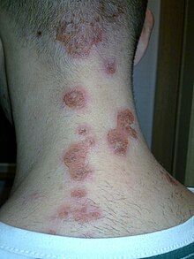
Chronic streptoderma is characterized by a relapsing course and the development of large (5-10 cm in diameter) foci of skin lesions. The foci are clearly limited spots with uneven, scalloped edges and a stratum corneum peeling off at the edges; most often they are localized on the legs. Bubbles appear on the skin, after opening which, large crusts of a yellowish-brown color are formed. After removal, a bright pink erosion is found at the site of the crust, from the surface of which serous-purulent exudate is abundantly separated. Between relapses, the formation of new bubbles ceases, instead of crusts, foci of peeling with gray-yellow scales form.
The prolonged existence of the infectious focus, as well as the increased sensitivity of the skin to microbes as a result of this, can lead to the transition of the disease from chronic streptoderma to microbial eczema . Characteristic signs of this process are the appearance of eczematous wells, a change in the boundaries of the lesion foci (with eczema, they become uneven, vague)